Image of the Month: Bovine endothelial cells dividing
The image is an example of the cutting-edge imaging and image analysis tools offered by Baylor’s Optical Imaging & Vital Microscopy Core.
Read More
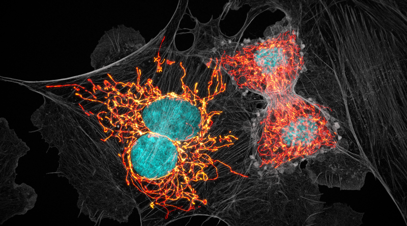
The image is an example of the cutting-edge imaging and image analysis tools offered by Baylor’s Optical Imaging & Vital Microscopy Core.
Read More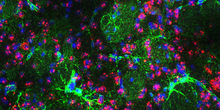
This confocal microscopy image shows a section of the brain of a mouse model of Huntington’s disease, with the astrocytes in green and cell nuclei in blue.
Read More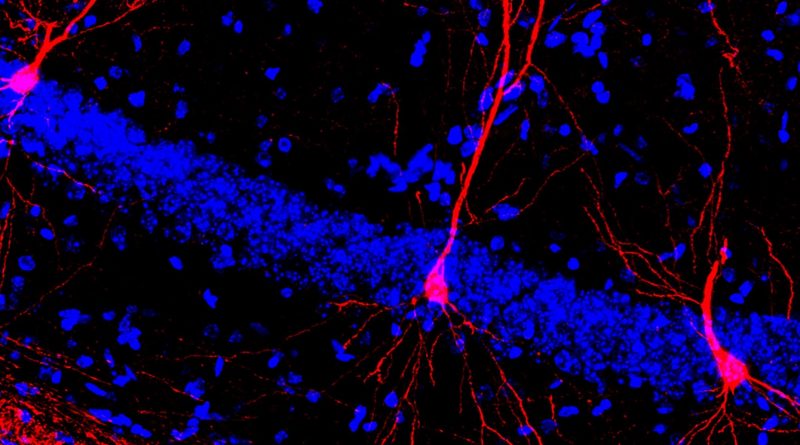
Early physical training triggers more dendritic arbors or cellular projections in hippocampal neurons (red), enhancing their functionality.
Read More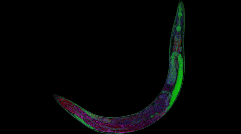
What is transparent, as long as a credit card is thick and helps solve science mysteries? The laboratory worm, C. elegans.
Read More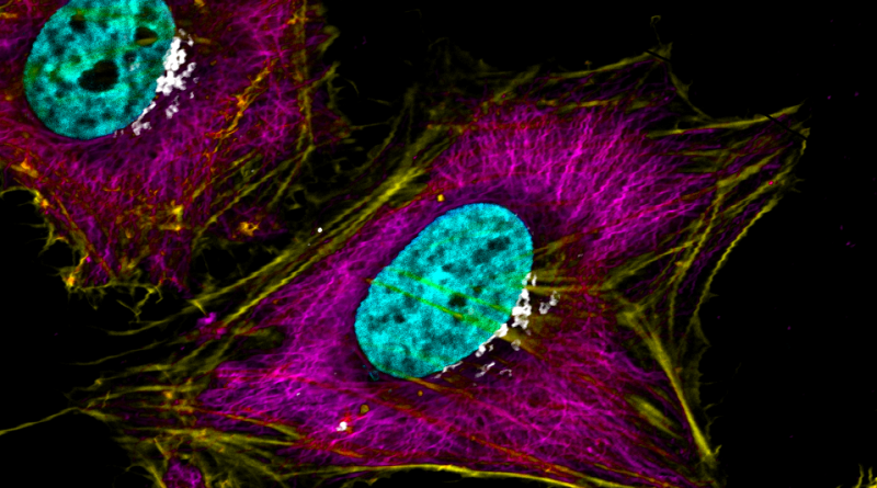
Our own Dr. Kristen Engevik is the winner of “CELL-ebrating HeLa” image competition with the image “Hues of HeLa.”
Read More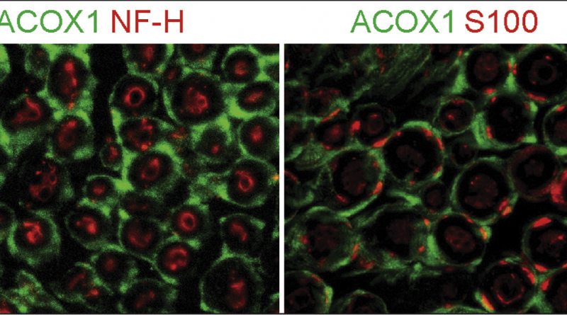
‘Transverse view of the sciatic nerve’ placed on top as the most viewed Image of the Month in 2020.
Read More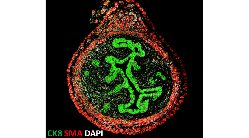
The image shows a cross-section of a uterus from a 3-week-old mouse.
Read More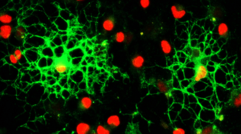
Oligodendrocytes produce and assemble myelin sheaths around nerves in the body to maintain rapid and precise neural communication.
Read More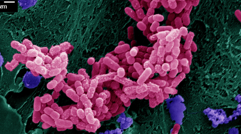
Infectious microorganisms, including disease-causing E. coli bacteria (pink) shown in the image, cause a third of all deaths world-wide.
Read More
Cancer cell clusters are more effective than single cells at spreading cancer in the body, and researchers at Baylor College of Medicine found out why.
Read More