Most viewed 2025 Image of the Month
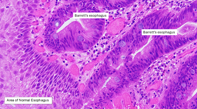
This microscopy view of the condition shows that an affected esophagus may show both normal tissue and diseased tissue with distinctive cellular characteristics.
Read More
This microscopy view of the condition shows that an affected esophagus may show both normal tissue and diseased tissue with distinctive cellular characteristics.
Read More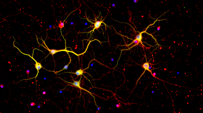
Dr. Juan Botas’s lab integrates laboratory, animal model and computational approaches to gain a deeper understanding of Huntington’s disease and other neurological disorders.
Read More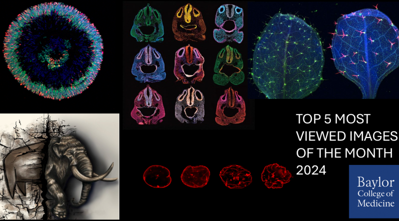
Our viewers selected these Images of the Month as the most popular of 2024.
Read More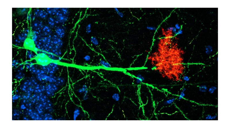
Neurons (green) and astrocytes (red) work together to regulate storage and retrieval of memories.
Read More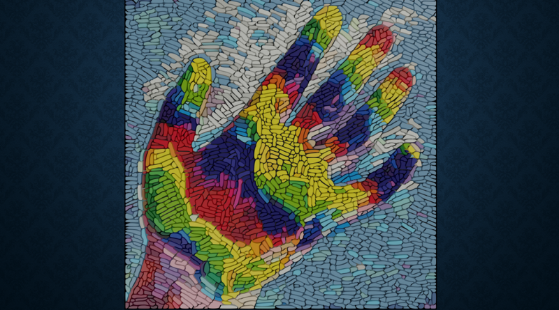
“We are all walking mosaics of genetically distinct cells,” said Dr. Margaret Goodell.
Read More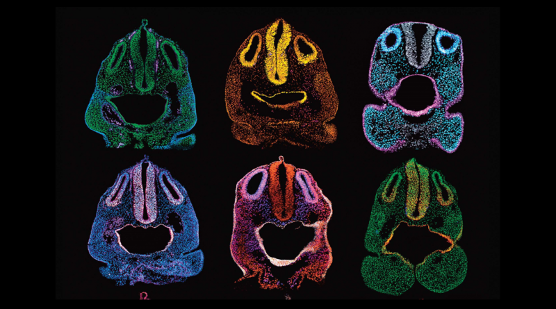
Sometimes, research inspires scientists like Dr. Ankita Thawani to dig into their artistic side to create renditions of their findings.
Read More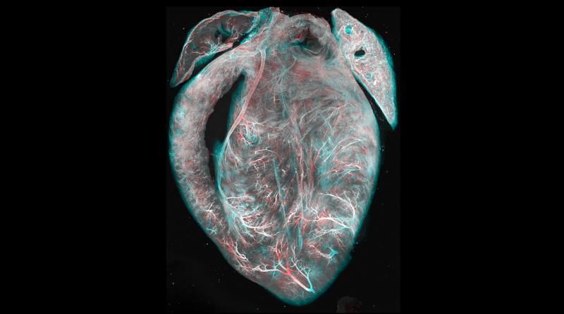
An improved tissue clearing protocol called EZ Clear renders entire organs, like this mouse heart, optically transparent for imaging.
Read More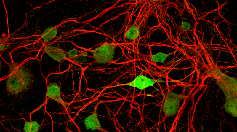
Like an elaborate superhighway, an intricate network of neuronal projections keeps neurons in the visual pathway connected.
Read More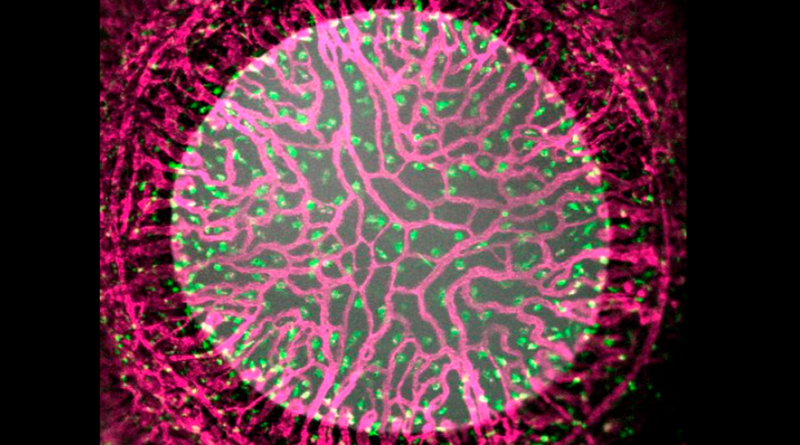
Microscopic details of a newborn mouse eye showing blood vessels (pink) and macrophage immune cells (green).
Read More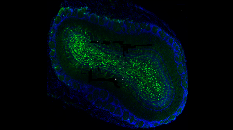
Oxytocin drives the development and synaptic integration of new neurons within the adult mouse brain contributing to brain plasticity.
Read More