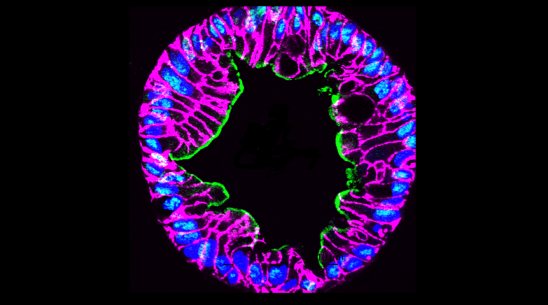
Image of the Month: Human intestinal enteroids
Human intestinal enteroids have revolutionized the study of gastrointestinal viruses like rotavirus.
Read More
Human intestinal enteroids have revolutionized the study of gastrointestinal viruses like rotavirus.
Read More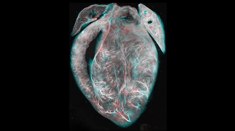
An improved tissue clearing protocol called EZ Clear renders entire organs, like this mouse heart, optically transparent for imaging.
Read More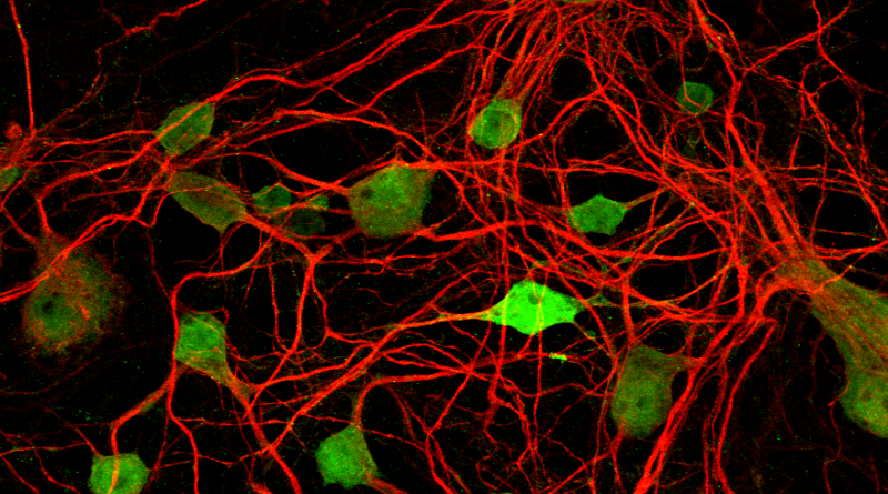
Like an elaborate superhighway, an intricate network of neuronal projections keeps neurons in the visual pathway connected.
Read More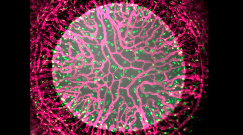
Microscopic details of a newborn mouse eye showing blood vessels (pink) and macrophage immune cells (green).
Read More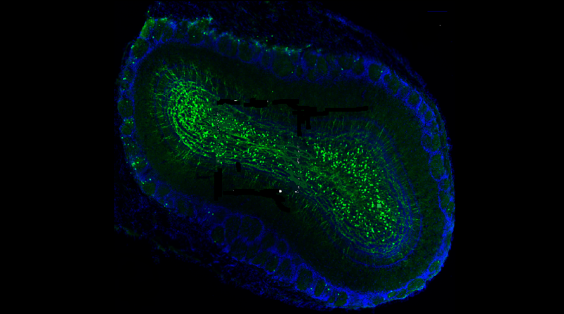
Oxytocin drives the development and synaptic integration of new neurons within the adult mouse brain contributing to brain plasticity.
Read More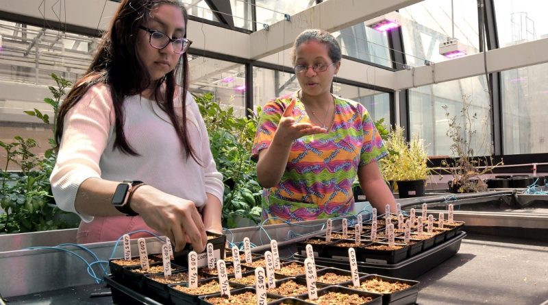
In addition to conducting groundbreaking biomedical research, Baylor College of Medicine is also creating better foods to address global food security issues.
Read More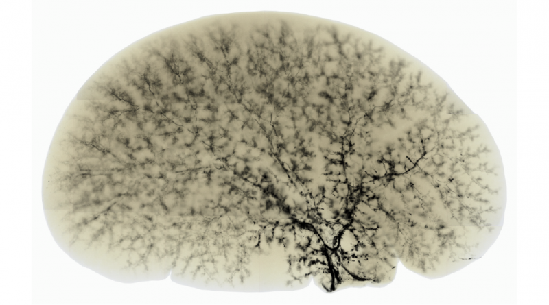
A healthy mouse liver typically shows a well-developed biliary tree through which bile flows into the intestine to help absorb fats and eliminate toxins.
Read More
From the Labs celebrates Heart Month with an image of a transparent mouse heart.
Read More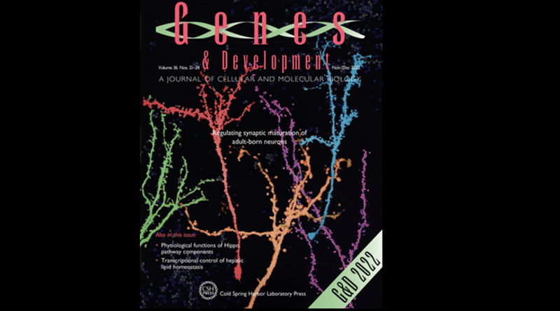
Congratulations to Brandon Pekarek, Benjamin Arenkiel and colleagues for making the cover of Genes & Development!
Read More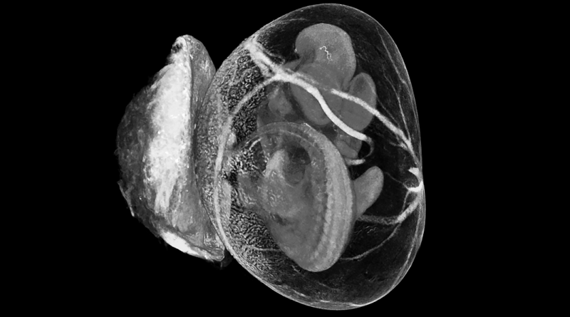
Using micro-computed tomography, Dr. Momal Sharif investigates structural defects within the mouse embryo, yolk sac and placenta.
Read More