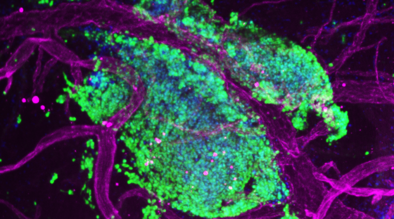
Image of the Month: A closer look at metastatic medulloblastoma
The image captures metastatic medulloblastoma tumor cells (green) intertwined with blood vessels (magenta) in the leptomeninges of a mouse model of the human condition.
Read More
The image captures metastatic medulloblastoma tumor cells (green) intertwined with blood vessels (magenta) in the leptomeninges of a mouse model of the human condition.
Read More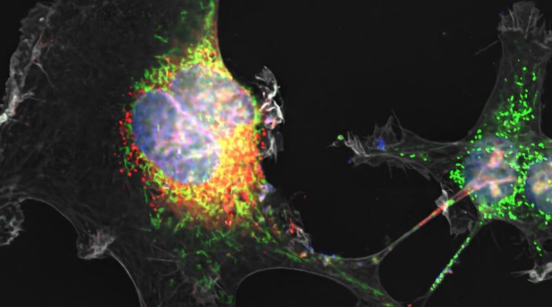
This is the first time that tunneling has been shown by Zika virus infection in placental cells.
Read More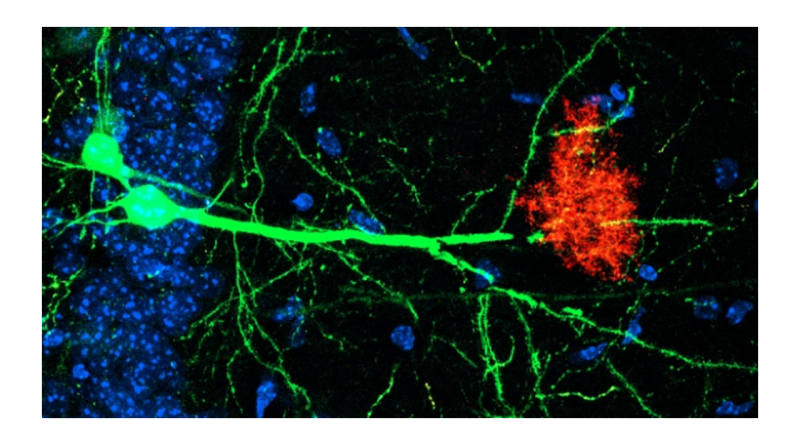
Neurons (green) and astrocytes (red) work together to regulate storage and retrieval of memories.
Read More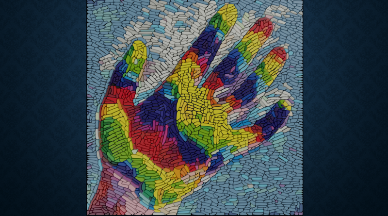
“We are all walking mosaics of genetically distinct cells,” said Dr. Margaret Goodell.
Read More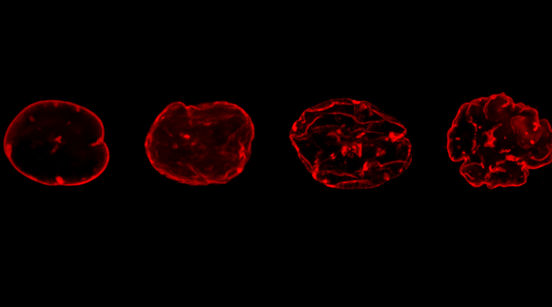
Overexpression of lamin A (red) causes structural changes to the cell nucleus that are associated with aging.
Read More
Researchers have peered into the chromosomes of the wooly mammoth, a magnificent creature that has been extinct for nearly 4,000 years.
Read More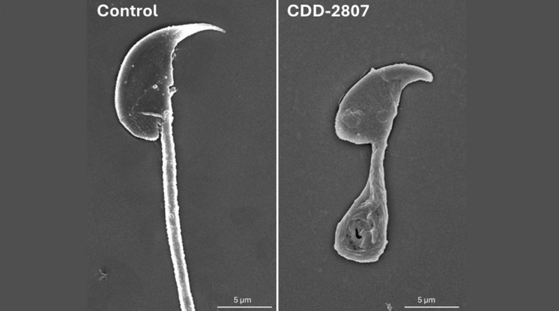
Alterations in normal sperm shape and motility led to infertility in an animal model and inspired work to develop a reversible birth control pill for men.
Read More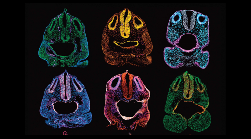
Sometimes, research inspires scientists like Dr. Ankita Thawani to dig into their artistic side to create renditions of their findings.
Read More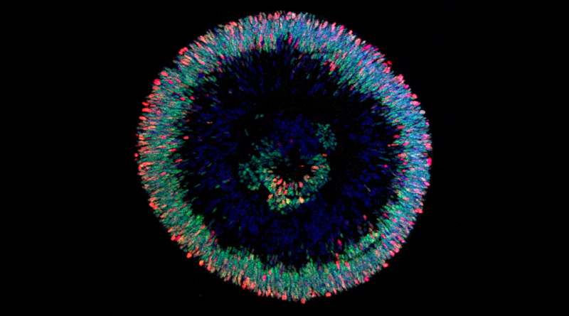
Retinal organoids are helping researchers study human retinal development and eye diseases such as glaucoma and macular degeneration.
Read More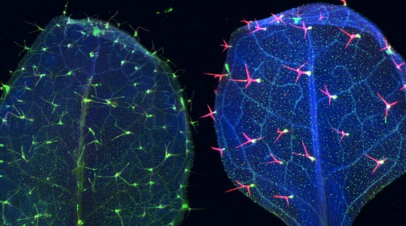
A serendipitous finding set researchers on a path to discover a potential way to make plants more flood tolerant.
Read More