Image of the Month: Human intestinal enteroids
Human intestinal enteroids (HIEs), like the one shown in this image, have revolutionized the study of gastrointestinal (GI) viruses like rotavirus.
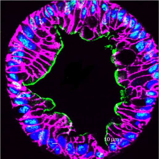
This is a confocal microscopy image of a human intestinal enteroid, showing the cell adhesion protein E-cadherin (magenta), sucrase isomaltase (green) – the intestinal enzyme on the surface of enterocytes that breaks down certain sugars and the cells’ nuclei detected by DAPI (blue). The image is courtesy of Drs. S. Crawford, H. Smith and M. Estes/PNAS, 2023.
These multicellular, non-transformed cell cultures retain host genetic properties, cellular organization and recapitulate the function of the human gastrointestinal epithelium. They serve as biologically relevant model systems for studying human gastrointestinal infections. HIE are one of many types of ‘organoids,’ which can be derived from normal adult tissues as well as diseased tissues.
Learn more about the research conducted in the Estes lab, here.
Follow From the Labs on Instagram!