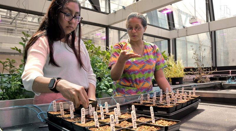
Image of the Month: The greenhouse inside the lab
In addition to conducting groundbreaking biomedical research, Baylor College of Medicine is also creating better foods to address global food security issues.
Read More
In addition to conducting groundbreaking biomedical research, Baylor College of Medicine is also creating better foods to address global food security issues.
Read More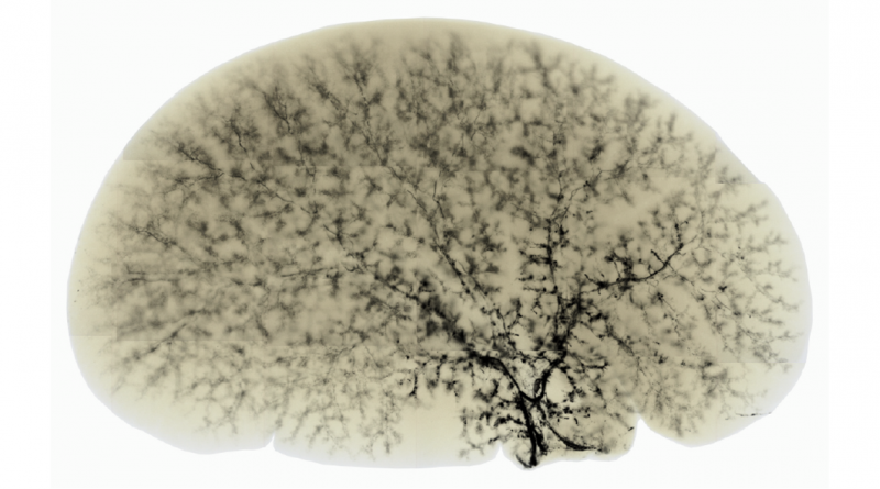
A healthy mouse liver typically shows a well-developed biliary tree through which bile flows into the intestine to help absorb fats and eliminate toxins.
Read More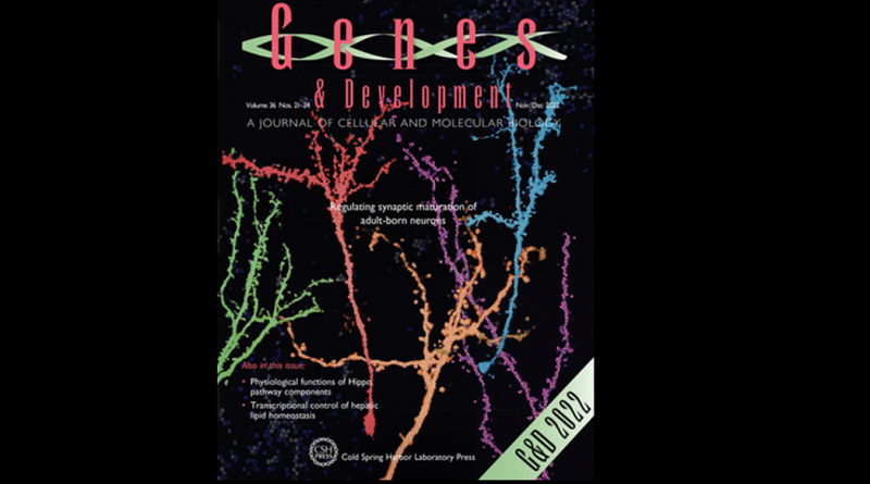
Congratulations to Brandon Pekarek, Benjamin Arenkiel and colleagues for making the cover of Genes & Development!
Read More
For years after the first one was posted, we have continued to feature amazing research images from Baylor labs every month.
Read More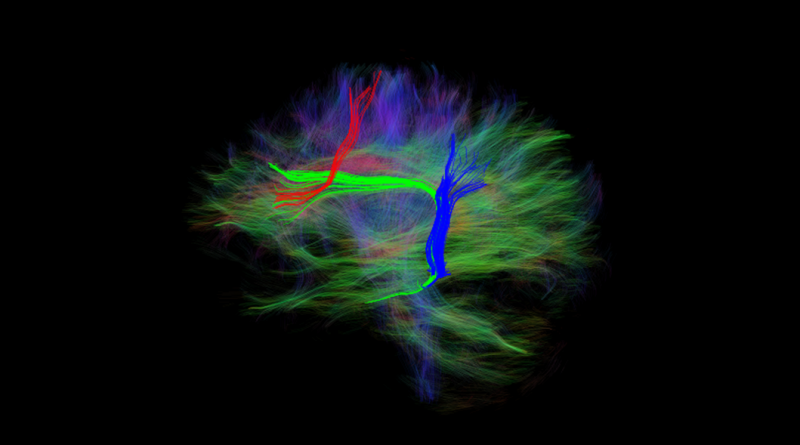
This work helps predict the types of brain damage and behavior that lead to chronic language loss, thus informing both psychological theories and treatment approaches to language.
Read More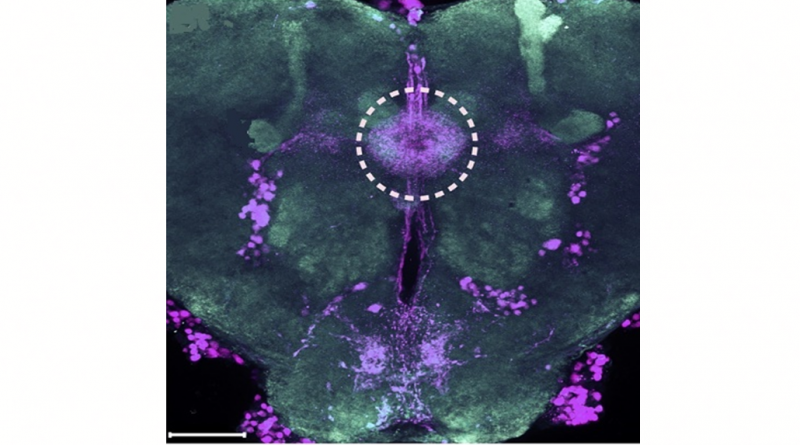
The image represents a visual perspective of a more efficient technology to study gene function in the laboratory fruit fly.
Read More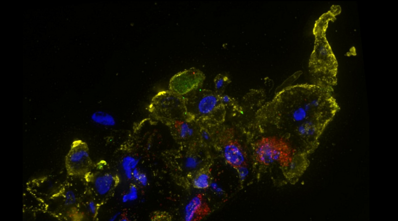
Immunofluorescence staining of human nose organoid cultures infected with SARS-CoV-2, the virus that causes COVID-19.
Read More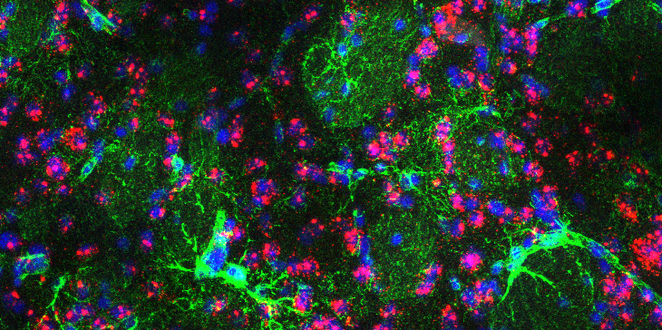
This confocal microscopy image shows a section of the brain of a mouse model of Huntington’s disease, with the astrocytes in green and cell nuclei in blue.
Read More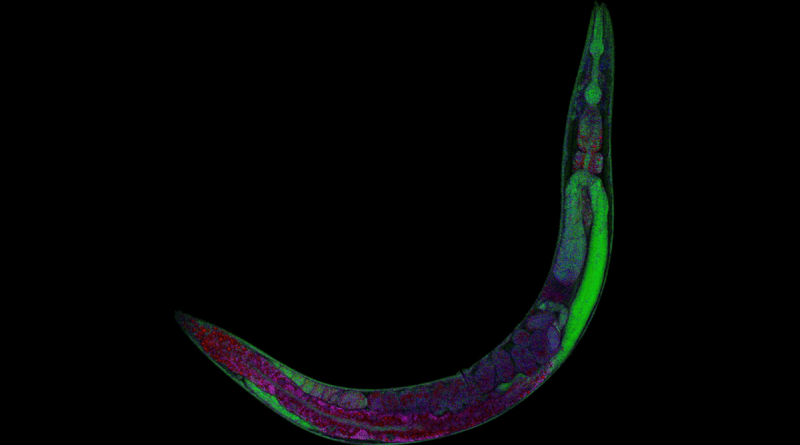
What is transparent, as long as a credit card is thick and helps solve science mysteries? The laboratory worm, C. elegans.
Read More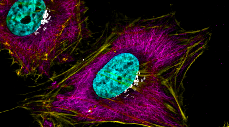
Our own Dr. Kristen Engevik is the winner of “CELL-ebrating HeLa” image competition with the image “Hues of HeLa.”
Read More