New imaging tools to listen in on the brain’s ‘electrical whispers’
Researchers at Baylor College of Medicine and colleagues at collaborating institutions have developed new biosensors to detect some of the smallest electrical signals that brain cells use to communicate. As reported in Nature Methods, these tools enable researchers to optically observe electrical activity in the brain with unprecedented sensitivity, across many cells and in deep regions of living animals – something that was previously impossible. These tools open new possibilities for studying how neural circuits process information.

“Neurons listen to many small electrical inputs –‘electrical whispers’– from other neurons. These small signals can be filtered, attenuated or amplified in the neuron. Depending on how these small electrical inputs combine, a neuron may respond with its own large spike of electrical activity. This process forms the basis for neuronal computation,” said corresponding author Dr. François St-Pierre, Vivian L. Smith Endowed Professor in Neuroscience, McNair Scholar and associate professor of biochemistry and molecular pharmacology at Baylor.

“To understand how the brain works in health and disease, we need to be able to study this electrical communication,” said Dr. Michelle A. Land, postdoctoral associate in neuroscience in the St-Pierre lab. “Researchers have already developed technologies that allow us to track and study the large spikes of electrical activity. But being able to monitor electrical whispers – also called subthreshold signals – is challenging with currently available optical tools. This study showcases new tools we developed to solve this problem.”
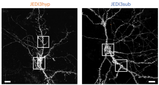
The researchers engineered two biosensors, JEDI3sub and JEDI3hyp. These sensors flash tiny lights inside neurons, changing brightness in response to electrical activity. Scientists can then use advanced microscopy techniques to track neuronal signals in real time.
A key advantage is that the new tools are designed to work well with two-photon microscopy, the preferred technique to image deep inside the brain.
“Earlier voltage sensors struggled to detect the millivolt-scale signals associated with subthreshold activity under two-photon microscopy. The new JEDI3 sensors were specifically designed to overcome this limitation,” Land said. “Using high-throughput screening, we tested hundreds of protein variants and selected two that produced stronger and clearer signals while remaining stable and bright.”
Compared with earlier tools, the new sensors produced signals that are more than three times as strong when cells experienced small voltage changes. The enhanced sensitivity to subthreshold voltage changes now allows researchers to listen in on the whispers of neurons.
Testing the new sensors in living mice
“We found that JEDI3sub could monitor subthreshold voltage activity in more than 100 neurons at once, something that was not possible before with this level of detail,” Land said. “This allowed us to see how groups of neurons fluctuate together and respond to sensory input, such as visual stimuli.
We discovered that most neurons in a given region showed coordinated patterns of activity, suggesting that the small voltage changes are shared across networks and may play a role in how the brain organizes information.”
The second sensor, JEDI3hyp, was particularly useful for tracking activity patterns linked to specific brain states. “For example, it captured subtle voltage changes in neurons during ‘sharp-wave ripples,’ brief bursts of coordinated brain activity associated with memory processing,” Land said. “It also revealed slow voltage changes that correlate with the animal’s internal state and level of alertness.” Changes in brain state can influence how the cell listens to the small electrical inputs from other neurons.
Importantly, the sensors worked not only in neuron cell bodies but also in fine structures like dendrites – the branching extensions where neurons receive inputs or ‘listen’ – thus enabling the study of how signals are integrated within individual cells, a phenomenon that has been difficult to measure in living brains due to the dendrites’ small size. JEDI3sub has enabled such studies to investigate the role dendrites play in information processing, as seen in the recent Science paper by Noguchi A. et. al. titled “Parallel Independent Voltage Computing along Dendrites of CA3 Pyramidal Neurons.”
Cell type specificity
“Another major advantage is cell-type specificity,” Land said. “Because these sensors are genetically encoded, they can be placed in specific types of neurons. We found that each neuron type, and even subpopulations within that type, exhibit different changes in brain state. This level of detail can help scientists understand how diverse cell types contribute to brain function.”
“Overall, the JEDI3 sensors represent a significant advance in neuroscience tools,” St-Pierre said. “They allow researchers to capture electrical activity in the brain with unprecedented sensitivity, across many cells and experiments, using different types of microscopes and in deep brain regions of living animals.
By making previously undetectable signals visible, these tools open new possibilities for studying how neural circuits process information and potentially discover how these processes may go wrong in disease.”
Mario Galdamez, Vincent Villette, Jun Zhu, Xiaoyu Lu, Mate Marosi, Shuyuan Yang, Gregory Foran, Alex J. McDonald, Xiaoyu Dong, Elsayed Zaabout, Haixin Liu, Zhuohe Liu , Kevin L. Colbert, Shujuan Lai, Matthew Shorey, Anthony S. G. Lourdiane, Annick Ayon, Jonathan Bradley, Caroline Mailhes-Hamon, Ryan G. Natan, Jian Zhong, Ryan Kroeger, Robert G. Law, Noura Hakam, Cameron L. Smith, Ming Hu, Shanii Tabb, Brice Bathellier, Barna Dudok, Na Ji, Laurent Bourdieu and Jacob Reimer also contributed to this work. The authors are affiliated with one or more of the following institutions: Baylor College of Medicine, INSERM, University of California-Berkeley, Rice University, Institut Pasteur, IHU reConnect-Paris and Lawrence Berkeley National Laboratory.
For financial support for this work, see the publication.
Follow From the Labs on X, BlueSky and Instagram!