Image of the Month: Faces of embryo development

Science is hard work, but also has many moments of personal satisfaction and some are quite fun. Sometimes, research inspires scientists, like Dr. Ankita Thawani, to dig into their artistic side and create renditions of their findings that not only reveal important insights into their study, but also catch our attention. In this case, they also caught the interest of the editor of Developmental Dynamics.

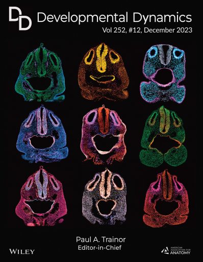
Thawani, a postdoctoral associate in Dr. Andy Groves lab, created this Image of the Month, which represents different sections of mouse embryos during development.
The story behind the image
From the Labs asked Thawani to tell the story behind this image.
FTL: What led you to take these images?
AT: As a new postdoc in Andy’s lab, I was acquainting myself with mouse embryonic morphology, so I collected serial sections throughout the embryo and systematically recorded them with fluorescent microscopy to construct an atlas for myself. When I reached the inner ear sections, I quickly noticed that they looked like an emotive face – the otic vesicles looked like ‘eyes,’ hindbrain like a ‘nose’ and pharyngeal arch made the ‘mouth.’
FTL: How did these initial images evolve into the artistic composition we see here, which made the cover of Developmental Dynamics?
AT: My first images were simple – only nuclear staining. As I collected more samples for experiments, the emotive face-like sections appeared to resemble different ‘expressions.’ In the next couple of years, as I performed varying combinations of immunostaining, I continued to store copies of the emoticon-looking images in a separate folder to make a journal cover submission to accompany my research paper. I chose the best from all the images I collected, pseudo-colored the immunostaining for artistic effect and constructed this rendition.
FTL: How does this image fit in your research?
AT: My research goal is to understand the genetic basis of vertebrate head development and how the brain, sensory organs, craniofacial skeleton and skin differentiate from the same sheet of cells. I think these colorful expressive heads are a fitting cover image to accompany my work.
Find the paper by Thawani, Groves et al that made the journal cover, here.

Dr. Andy Groves is the Vivian L. Smith Endowed Chair in Neuroscience and professor of molecular and human genetics at Baylor College of Medicine.
Follow From the Labs on X @BCMFromtheLabs and Instagram!