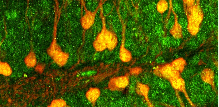
Image of the month: This brain region coordinates survival adaptations to food shortages
SRC-2 (green) in POMC neurons in the hypothalamus helps animals survive when food is hard to find.
Read More
SRC-2 (green) in POMC neurons in the hypothalamus helps animals survive when food is hard to find.
Read More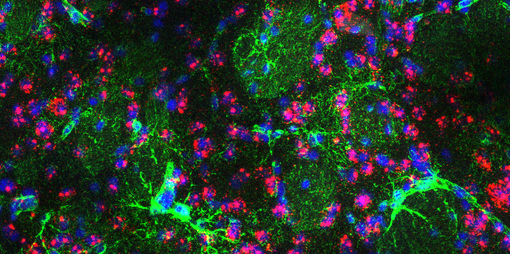
Brain of a mouse model of Huntington’s disease.
Read More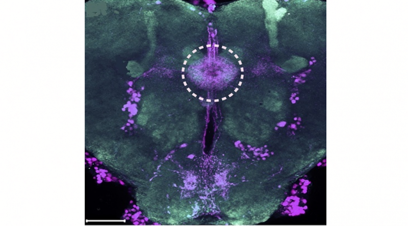
The image represents a visual perspective of a more efficient technology to study gene function in the laboratory fruit fly.
Read More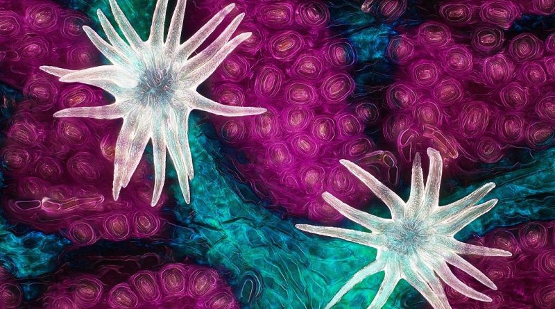
Three essential elements of plant life raised to the top of Nikon’s Small World Photomicrography Competition.
Read More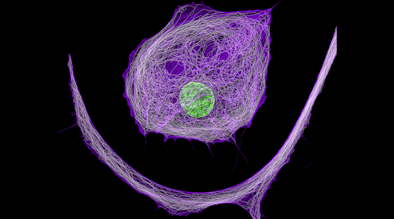
The Optical Imaging & Vital Microscopy Core Lab is dedicated to vital and intravital imaging of a broad range of biological processes.
Read More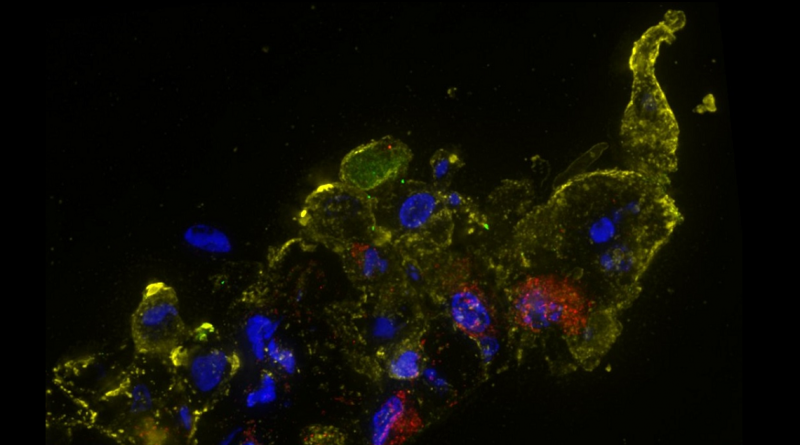
Immunofluorescence staining of human nose organoid cultures infected with SARS-CoV-2, the virus that causes COVID-19.
Read More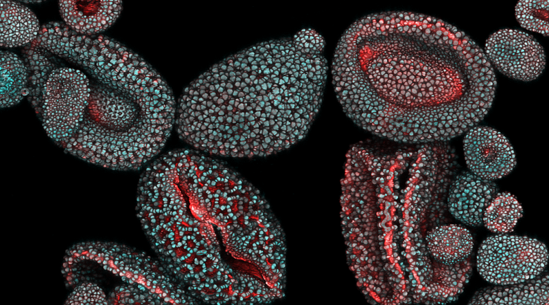
The image is an example of the cutting-edge imaging and image analysis tools offered by Baylor’s Optical Imaging & Vital Microscopy Core.
Read More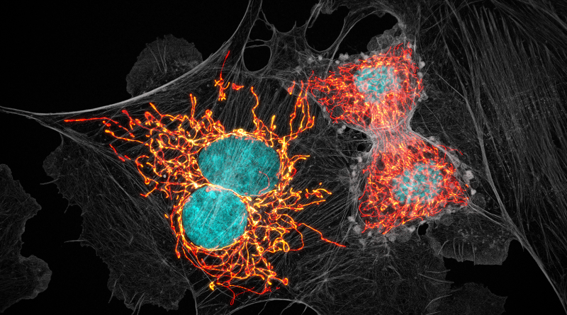
The image is an example of the cutting-edge imaging and image analysis tools offered by Baylor’s Optical Imaging & Vital Microscopy Core.
Read More
This confocal microscopy image shows a section of the brain of a mouse model of Huntington’s disease, with the astrocytes in green and cell nuclei in blue.
Read More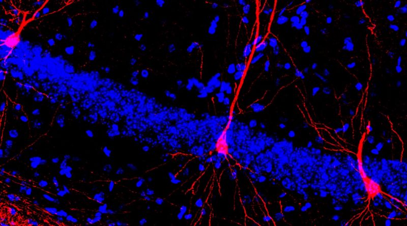
Early physical training triggers more dendritic arbors or cellular projections in hippocampal neurons (red), enhancing their functionality.
Read More