Glial cells help mitigate neurological damage in Huntington’s disease
Research with human samples and mouse and fruit fly models opens a window into the brain’s protective responses in Huntington’s disease.
Read More
Research with human samples and mouse and fruit fly models opens a window into the brain’s protective responses in Huntington’s disease.
Read More
One day, people might be able to control diabetes from the brain.
Read More
Study reveals key regulatory mechanism for coinciding obesity and mental disorders and suggests the possibility of pharmacological treatment.
Read More
The findings support that an altered daily rhythm of expression of the Rev-erb gene in the brain may underlie dawn phenomenon in diabetes.
Read More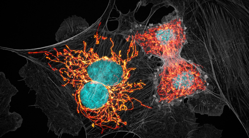
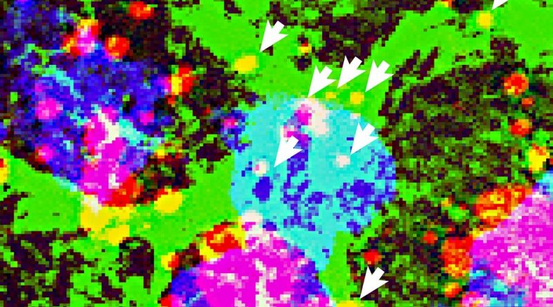
The image is an example of the cutting-edge imaging and image analysis tools offered by Baylor’s Optical Imaging & Vital Microscopy Core.
Read More
Health experts from the U.S. and France hosted the COVID-19 Disaster Research and Prevention Symposium to share what they have learned during the pandemic.
Read More
NANOS2-deficient germ cells failed to mature into sperm, remained pluripotent and were more likely to transform into embryonic cancer cells.
Read More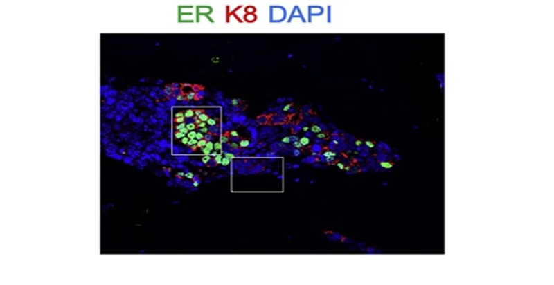
The bone microenvironment prompts changes in breast cancer cells that help them escape treatment and become more invasive.
Read More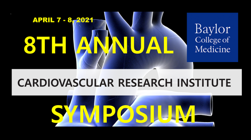
Review the symposium highlights, visit the virtual poster session and meet the winners of the first Dr. Mark L. Entman Award for Excellence in Cardiovascular Education.
Read More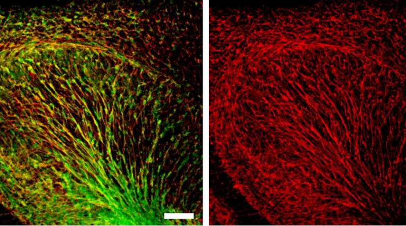
Membrane-bound BASP-1 protein is a possible biomarker of neural stem cells that would allow the study of adult human neurogenesis.
Read More