The Most Viewed Image of the Month in 2020
‘Transverse view of the sciatic nerve’ placed on top as the most viewed Image of the Month in 2020.
Read More
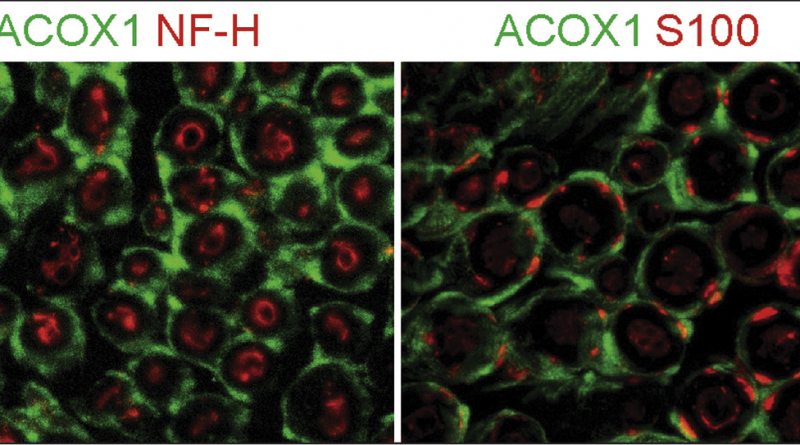
‘Transverse view of the sciatic nerve’ placed on top as the most viewed Image of the Month in 2020.
Read More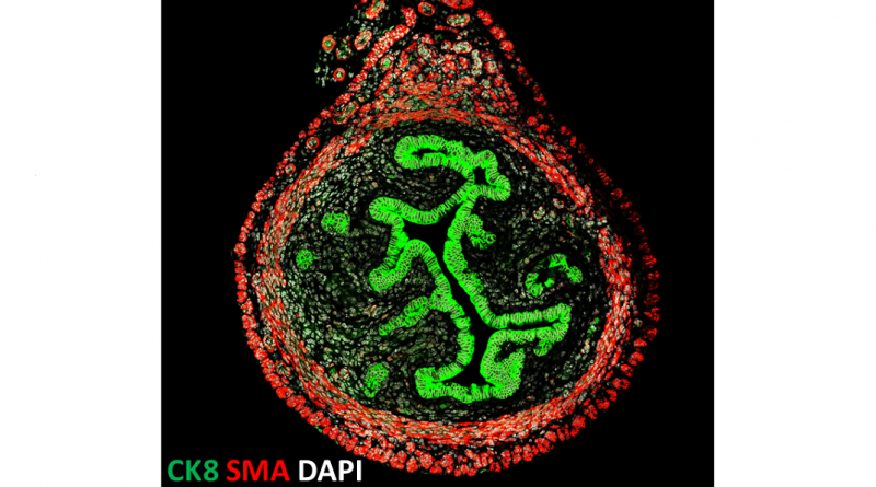
The image shows a cross-section of a uterus from a 3-week-old mouse.
Read More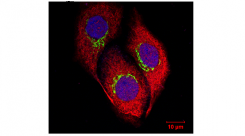
Pinpointing the location of Man1b1 inside the cell sharpened the view of cellular regulation of protein homeostasis.
Read More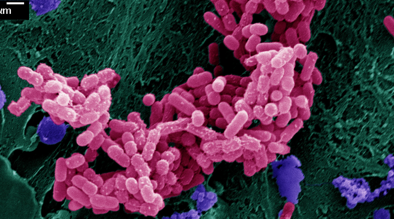
Infectious microorganisms, including disease-causing E. coli bacteria (pink) shown in the image, cause a third of all deaths world-wide.
Read More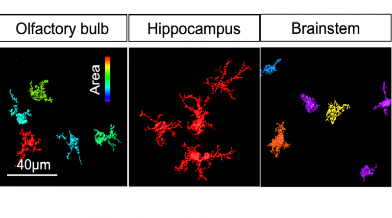
Astrocytes, the most abundant cells in the brain, have surprised researchers with their unanticipated diversity of shapes and functions. This month, From the Labs’s features
Read More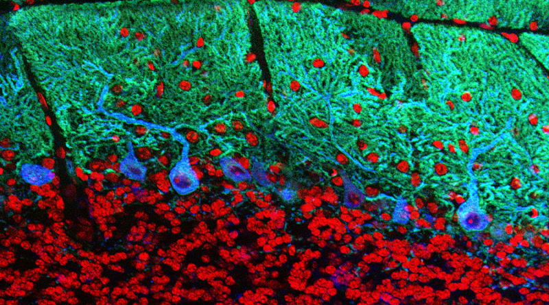
Purkinje cells are a type of neuron located in the cerebellar cortex of the brain. Purkinje cells are involved in the regulation of movement,
Read More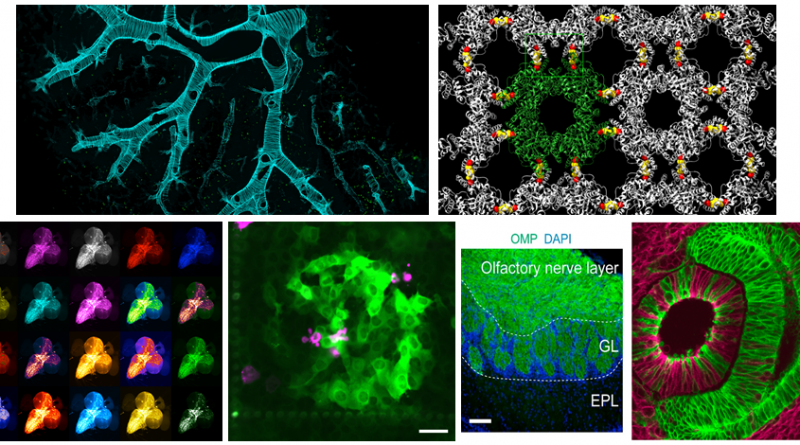
From the Labs wraps up the Image of the Month feature for the year by presenting a slide show with the top six most viewed
Read More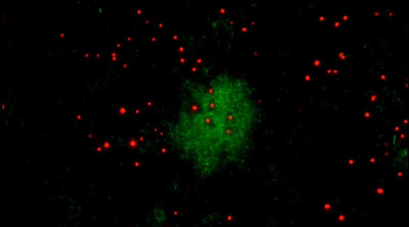
From the Labs presents the movies of the month, featuring the soil-dwelling single-celled amoeba Dictyostelium discoideum interacting with Gram-negative bacteria Klebsiella pneumoniae, courtesy of the lab
Read More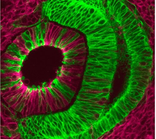
The Ross Poché lab is dedicated to elucidating the transcriptional and epigenetic mechanisms regulating proliferation and differentiation of retinal progenitor cells to identify new therapeutic
Read More
On the cover! The diversity of adult-born neurons. Different colors indicate different adult-born granule cell types and delineate their dendritic arborization
Read More