Connecting Tau aggregates, transposable elements and neurodegeneration in Alzheimer’s disease
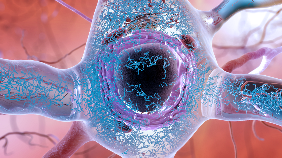
Although it’s been known for years that the accumulation of Tau protein within brain cells in combination with progressive cell death is one of the key characteristics of Alzheimer’s disease, it still is not clear why brain cells ultimately die.
In the Integrative Functional Genomics Lab at Baylor College of Medicine, Dr. Joshua Shulman and his colleagues integrate human genomic analyses with functional investigation in fruit fly models of neurodegenerative disease to better understand the genetic mechanisms of Alzheimer’s and Parkinson’s disease, and to translate these insights into enhanced risk prediction, diagnosis and treatment of these disorders.

In this study, the researchers investigated how accumulation of Tau protein may trigger neurodegeneration in Alzheimer’s disease.
“One thing that scientists have noticed over the years is that neurons affected by Tau accumulation also appear to have genomic instability,” said Shulman, associate professor of neurology, neuroscience and molecular and human genetics at Baylor College of Medicine and investigator at the Jan and Dan Duncan Neurological Research Institute at Texas Children’s Hospital.
Genomic instability refers to an increased tendency to have alterations in the DNA, such as mutations or other impairments that result in the genome not functioning correctly.
“Genomic instability is known to be a major driving force behind other diseases such as cancer,” Shulman said. “Our study focused on a new possible causal connection between Tau accumulation within neurons and the resulting genomic instability in Alzheimer’s disease.”
Enter transposable elements
Previous studies of brain tissues from patients with other neurologic diseases and of animal models have suggested that neurons not only present with genomic instability, but also with activation of transposable elements.
“Transposable elements are short pieces of DNA that do not seem to contribute to the production of proteins that make cells function. They behave in a way similar to viruses; they can make copies of themselves that are inserted within the genome and this can create mutations that lead to disease,” Shulman said. “Although most transposable elements are dormant or dysfunctional, some may become active in human brains late in life or in disease. That’s what led us to look specifically at Alzheimer’s disease and the possible association between Tau accumulation and activated transposable elements.”
Shulman and his colleagues began their investigations by studying more than 600 human brains from a population study run by co-author Dr. David Bennett at Rush University Medical Center in Chicago. This population study follows participants throughout their lives and at death, allowing the researchers to examine their brains in detail postmortem. One of the evaluations is the amount of Tau accumulation across many brain regions. In addition, co-author Dr. Philip De Jager at the Broad Institute and Columbia University comprehensively profiled gene expression in the same brains.

“With this large amount of data, we looked to identify signatures of active transposable elements, but this was not easy,” Shulman said. “We therefore reached out to Dr. Zhandong Liu, a co-author in this study, and together we developed a new software tool to detect signatures of active transposable elements from postmortem human brains. Then we conducted a statistical analysis in which we compared the amount of active transposable elements signatures with the amount of Tau accumulation, brain by brain.” Liu also is assistant professor of pediatrics – neurology at Baylor and a member of the Dan L Duncan Comprehensive Cancer Center.
The researchers found the first evidence of a strong link between the amount of Tau accumulation in neurons and detectable activity of transposable elements.
“We identified individual transposable elements that were active when Tau aggregates were present. Surprisingly, we also found evidence that the activation of transposable elements was quite broad across the genome,” Shulman said.
Other research has shown that Tau may disrupt the tightly packed architecture of the genome. It is believed that tightly packed DNA limits gene activation, while opening up the DNA may promote it. Keeping the DNA tightly packed may be an important mechanism to suppress the activity of transposable elements that lead to disease.
“The fact that Tau aggregates can affect that architecture of the genome may be one possible mechanism by which transposable elements are activated in Alzheimer’s disease,” Shulman said. “However, our studies in human brains only establish an association between Tau accumulation and activation of transposable elements. To determine whether Tau accumulation could in fact cause transposable element activation, we conducted studies with a fruit fly model of Alzheimer’s disease.”
In this fruit fly model of the disease, the researchers found that triggering Tau changes similar to those observed in human brains resulted in the activation of fruit fly transposable elements, strongly suggesting that Tau aggregates that disrupt the architecture of the genome can potentially mediate the activation of transposable elements and ultimately cause neurodegeneration.
“We think our experiments reveal new and potentially important insights relevant for understanding Alzheimer’s disease mechanisms,” Shulman said. “There is still a lot of work to be done, but by presenting our results we hope we can stimulate others in the research community to help work on this problem.”
Find all the details of this work in the journal Cell Reports.
Other key contributors to this work include Caiwei Guo, Hyun-Hwan Jeong, Yi-Chen Hsieh at Baylor College of Medicine and Texas Children’s Hospital, and Hans-Ulrich Klein at Columbia University Medical Center and the Broad Institute.
This study was supported by grants from the National Institutes of Health (R01AG053960, R01AG050631, U01AG046161, R01AG033193, C06RR029965, P30AG10161, R01AG15819, R01AG17917, U01AG46152, R01AG36836, R01GM120033, R01AG36042 and RC236547), the Alzheimer’s Association and the American Federation for Aging Research. This work was also supported by the Huffington Foundation, Jan and Dan Duncan Neurological Research Institute at Texas Children’s Hospital, the Burroughs Wellcome Fund, the National Science Foundation (DMS-1263932), Cancer Prevention Research Institute of Texas (RP170387), Houston Endowment and the Belfer Neurodegenerative Disease Consortium.