Image of the Month: Tau travels between neurons
Tau-containing molecular aggregates inside neurons are the defining features of Alzheimer’s disease (AD). At Baylor College of Medicine, Dr. Cristian Lasagna-Reeves‘ lab focuses on elucidating the role that tau physiology plays in AD and in more than 20 other neurodegenerative tau-caused conditions.
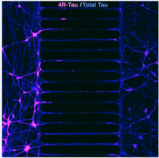
The image shows two distinct human neuronal populations in separate compartments of a microfluidic device. On the left, neurons express the 4R tau form (pink), which is associated with toxicity in Alzheimer’s disease, while on the right there are neurons from healthy individuals expressing normal tau (blue). Across the central microgroove region of the device, neurons extend axons between the compartments. From the Labs thanks Dr. Nur Jury-Garfe, neurology instructor in the Lasagna-Reeves lab for providing the image.
“These microfluidic platforms are particularly powerful in Alzheimer’s disease research because they allow us to physically isolate neuronal populations while still studying axonal connectivity and tau propagation across compartments,” Jury-Garfe explained.


Follow From the Labs on X, BlueSky and Instagram!