Simple, rapid and robust method makes mouse whole organs transparent for imaging
The adage ‘Seeing is believing’ was front of mind for Dr. Chih-Wei Logan Hsu and Dr. Joshua D. Wythe at Baylor College of Medicine as they and their colleagues developed an innovative technology called EZ Clear. This new tissue clearing method has simplified and sped up the process to render tissue optically transparent, which enables 3D imaging of entire, intact tissues or even entire organs. Their new method, published in the journal eLife, was developed at the Optical Imaging and Vital Microscopy Core (OiVM) at Baylor.
Tissue clearing and whole organ imaging have revolutionized biology, enabling the exploration of organs in three-dimensional space without compromising tissue architecture.

“Previous methods were complicated, laborious and often required expensive equipment, as well as the use of hazardous organic solvents, all of which prevented the widespread adoption of these methods,” said Hsu, co-director of OiVM and assistant professor of integrative physiology and education, innovation and technology at Baylor. “These difficulties motivated us to develop a simpler clearing process that users could more easily complete, saving time and valuable resources to focus on the actual questions they want to investigate in their systems.”

“The beauty of this method is that you can analyze the sample from a global or macro view without physically disturbing the natural organization of the tissue or organ,” said Wythe, associate professor of integrative physiology and of neurosurgery at Baylor. He also is a member of the Dan L Duncan Comprehensive Cancer Center and the Cardiovascular Research Institute.
“For instance, researchers can now visualize neuronal connections between the eye and the brain. If done in sections, the process would disturb the natural organization of the tissues and is incredibly difficult to reconstruct in 3D, limiting our understanding of the connections between neurons and other surrounding cells over larger volumes or areas. 3D imaging circumvents these limitations, and the advent of EZ Clear makes 3D imaging accessible to most modern molecular biology laboratories,” Wythe said.
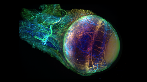
A mouse eye rendered transparent with EZ clear and imaged on Zeiss Lightsheet Z. Video created by Dr. C.-H. L. Hsu.
EZ Clear also preserves endogenous and synthetic labeling methods, such as fluorescence, without altering sample size.
The study shows successful clearing and labeling of neurons and blood vessels within the brain, as well as vessels within the eye, heart, kidney, testis and ovary, and successful whole organ clearing of mouse lung, liver and pancreas.
“EZ Clear eliminates previous technical barriers, allowing researchers to inspect their organs of interest from a macro, whole organ level, down to a cellular resolution,” Wythe said. “It has eliminated previous practical, safety and economical challenges, while providing reproducible, high-quality whole organ visualization, which is an important perspective as it can provide new insights on the topic being investigated.”
“Researchers can now learn this easier-to-complete, reproducible tissue clearing process at the OiVM. They can then implement it in their own labs and obtain results in 48 hours by following three simple steps, while other methods need weeks or even months to clear the tissue,” Hsu said. “Then, they can bring the cleared organ to the OiVM for 3D imaging and analysis.”
Other contributors to this work include Juan Cerda, Jason M. Kirk, Williamson D. Turner, Tara L. Rasmussen, Carlos P. Flores Suárez and Mary E. Dickinson. The authors are all affiliated with Baylor College of Medicine.
This project was supported by the Optical Imaging and Vital Microscopy Core and the Bioengineering Core at Baylor College of Medicine. Further support was provided by grants from the National Institutes of Health (5T32GM088129-10, R01HL146745, R01HD099026, U42OD026645, R01HL159159 and 1S10OD016167), the American Heart Association (22PRE916015), the Cancer Prevention Research Institute of Texas (RP200402), the Department of Defense (W81XWH18-1-0350) and the Canadian Institutes of Health Research (PJT-155922).