MiMIC proves versatile tool for fruit fly research
By Ruth SoRelle, M.P.H.
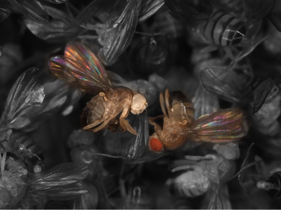
The “Swiss Army Knife” of the fruit fly laboratory – dubbed MiMIC (Minos-mediated integration cassette) – provided the basis for building a large protein collection that allows researchers to assess expression of genes and proteins under the microscope in unfixed tissues as well as to assess the consequences of knocking down protein activity in vivo, said researchers from Baylor College of Medicine and the Jan and Dan Duncan Neurological Research Institute at Texas Children’s Hospital and Baylor in a report that appears online in the journal eLife.
Tag proteins
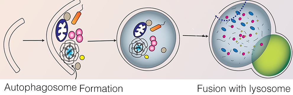
Dr. Hugo Bellen, professor of molecular and human genetics and director of the Program in Developmental Biology at Baylor said that when the first experiments were done with MiMIC in 2011, it was a proof of principle. They could “tag” proteins with a green fluorescent protein using transposon insertions that integrated in the middle of genes.
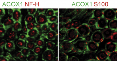
“In this report we describe the generation of an additional 6,000 MiMICs,” said Bellen. “We tagged nearly 400 different genes with a green fluorescent protein (GFP). These tagged genes are extremely useful because you can monitor GFP expression and do live imaging. That means that we can see the fluorescent protein under a microscope. For 90 percent of the genes, we can look under the scope and see the expression and distribution of the protein – no staining or other manipulations are needed.”
Important methodology

In this paper, the researchers with first authors Sonal Ngarkar-Jaiswal, Pei-Tseng Lee and Megan E. Campbell, post-doctoral associates in Bellen’s lab at Baylor, also found that they could degrade the GFP- tagged proteins to produce lower protein levels in a tissue-specific manner and then reverse the protein loss. This allows them to determine if the gene is associated with specific symptoms in specific cells and if the symptoms can be reversed (phenotype).
Eventually, Bellen said, he hopes to tag 5,000 genes by collaborating with other colleagues in Harvard, Carnegie, Toronto and Leuven to evaluate expression patterns and associated phenotypes.
Others who took part in this work include Kuchuan Chen, Stephanie Anguiano-Zarate, Manuel Cantu Gutierrez, Theodore Busby, Wen-Wen Lin, Yuchun He, Karen L. Schulze, Koen J.T. Venken, all of Baylor; Robert W. Levis and Allan C. Spradling of Carnegie Institution for Science in Baltimore, Maryland; Roger A. Hoskins, Benjamin W. Booth and Martha Evans-Holm of Lawrence Berkeley National Laboratory in Berkeley, California.
Funding for this work came from National Institutes of Health/National Institute of General Medical Science (Grant R01GM067858 to H.J.B., 3R01GM067858-11S1 to S.A., 3R01GM067858-09S1 to T.B.), the Robert A. and Renée E. Belfer Family Foundation, Baylor College of Medicine, the McNair Medical Institute, the March of Dimes Foundation (Grant #1-FY14-315), the Cancer Prevention and Research Institute of Texas (Grant R1313), and the National Institutes of Health (Grant1R21GM110190). Confocal microscopy was supported by NICHD P30HD024064 to the Baylor College of Medicine Intellectual and Developmental Disabilities Research Center.