Image of the Month: A closer look at metastatic medulloblastoma
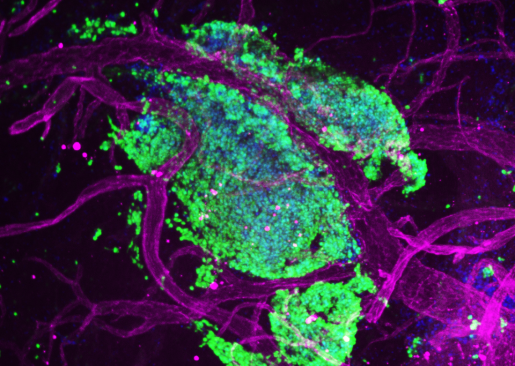
When medulloblastoma cells metastasize or spread away from their original tumor site, they invade the leptomeninges, protective layers surrounding the brain and the spinal cord. The image this month captures a niche of metastatic medulloblastoma tumor cells (green) intertwined with blood vessels (magenta) in the leptomeninges of a mouse model of the human condition.
Metastases are the most common and most important cause of illness and death for children with medulloblastoma, the most prevalent malignant pediatric brain tumor. Drs. Namal Abeysundara, Michael D. Taylor and their colleagues at Baylor College of Medicine, Texas Children’s Hospital and collaborating institutions wanted to better understand how these tumors spread and grow on the leptomeninges. They hoped the findings could potentially help them develop better treatments to improve survival and quality of life for affected children.
Their investigation revealed that metastatic medulloblastoma cells recruit local fibroblasts to back up tumor spread and growth. Disrupting this association could be a promising strategy for treating this devastating childhood condition.
Learn more about this research. Read the paper in Nature Cell Biology and an interview with the authors in From the Labs.

Dr. Namal Abeysundara is a postdoctoral fellow in Dr. Michael D. Taylor’s lab.
 Dr. Michael D. Taylor is a professor of pediatrics, section of hematology-oncology, and of neurosurgery at Baylor College of Medicine, and is a staff neurosurgeon in the Department of Neurosurgery at Texas Children’s Hospital. He also is the Cyvia and Melvyn Wolff Chair of Pediatric Neuro-Oncology at the Texas Children’s Cancer and Hematology Center and a member of Baylor’s Dan L Duncan Comprehensive Cancer Center.
Are you interested in the research conducted in Dr. Taylor’s lab? Visit their website.
Follow From the Labs on X, BlueSky and Instagram!