Like a mastermind of its own destiny, glioma sets a stage that favors its own growth
“In some regards, glioma brain tumors exhibit Machiavellian behavior,” said Dr. Benjamin Deneen. Machiavellian, as in scheming and cunning, comes from the Italian philosopher Niccolò Machiavelli who in the 1500s wrote about ‘the ends justify the means’ behavior, in particular among politicians.
Deneen, who is professor of neurosurgery and in the Center for Stem Cell and Regenerative Medicine at Baylor College of Medicine, was referring to the fascinating evidence he and his colleagues uncovered about how glioma manipulates its environment in ways that favor its own growth. This research, published in Nature, suggests new strategies to treat patients with this condition, one of the most aggressive malignant primary brain tumors.

Identifying gene variants that drive glioma
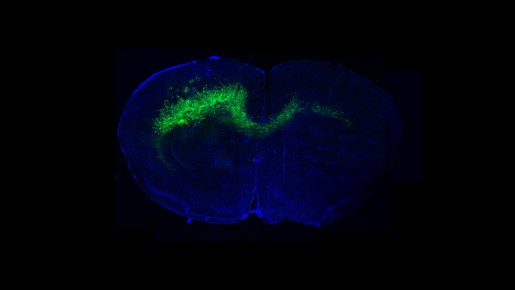
The original goal of this study was to develop an experimental system that would enable researchers to identify new cancer genes in mouse models of brain tumors. To achieve this goal, a collaboration began between the Deneen lab and Baylor co-author Dr. Kenneth L Scott. Together, they genetically engineered their mouse model of glioma into a novel, high-throughput screening platform to identify variants of the PIK3CA gene that drive tumor progression.
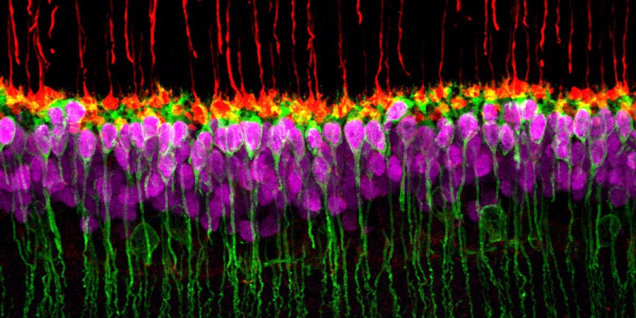
Using their novel screening platform, the researchers discovered several variants of PIK3CA that drive glioma development. Two of the PIK3CA variants, named C420R and H1047R, stood out because they were the strongest drivers of tumor development. Interestingly, some of the genes specifically expressed in C420R and H1047R gliomas are involved in synapse formation – the junctions between neurons through which they communicate – suggesting that the tumors may affect the synaptic balance of neighboring neurons.
“These gene variants produce proteins that differ in only one amino acid – the building blocks of proteins – yet some of the variants generate tumors with molecular profiles that are quite different from the others.
This was quite a surprise and told us that seemingly similar PIK3CA variants promote glioma formation through very different mechanisms,” said Deneen, who also is a member of the Dan L Duncan Comprehensive Cancer Center and holds the Marianne and Russell Blattner Chair at Baylor.
Glioma sets conditions that favor its own progression
To investigate these different mechanisms, Deneen and colleagues focused on the synaptic gene signatures, hypothesizing that these alterations in synaptic gene expression could lead to seizures, network hyperexcitability and direct synaptic changes in their mouse model of glioma. To conduct these studies, Deneen partnered with co-author Dr. Jeffrey L Noebels, professor of neurology, neuroscience, and molecular and human genetics and Cullen Trust for Health Care Endowed Chair in Neurogenetics at Baylor.
“It is well established that synaptic imbalance can result in extensive changes in neuronal network connectivity and excitability, which in some cases culminates in seizure activity,” Deneen said. “Seizures are typical in glioma, but the underlying cellular and genetic mechanisms are not well understood. We took this finding as an opportunity to explore whether different PIK3CA variants can induce epilepsy in glioma and also to understand more about the mechanisms by which tumors promote neuronal hyperexcitability.”
Their studies showed that, indeed, gliomas driven by C420R and H1047R variants do promote early onset of hyperexcitability in neurons surrounding the tumor and remodel synaptic networks by inducing synapse formation. Mice carrying these tumors had seizures that appeared much earlier than in mice bearing tumors driven by other PIK3CA variants.
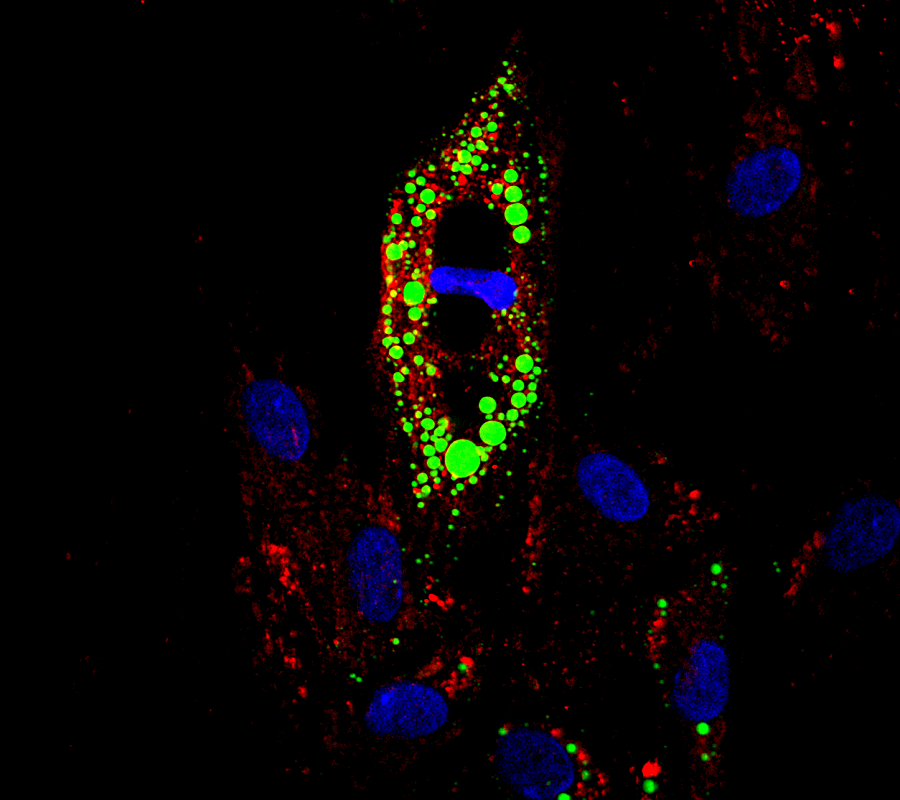
Digging deeper into the mechanisms that mediate the effect of C420R and H1047R gliomas on their microenvironment, the researchers discovered that these gliomas selectively secreted several molecules of the glypican (GPC) family and that GPC3 drove hyperexcitability and synaptic remodeling. Further, they found that GPC3 itself can drive glioma formation.
These findings provide the first evidence of a glioma-derived mechanism that manipulates the neuronal microenvironment during tumor progression.
“We have uncovered a central mechanism by which glioma alters neurons to establish environmental conditions in the brain that support growth. Therapeutically, we are actively examining how short circuiting glioma-to-neuron communication can be used to treat patients with these malicious brain tumors,” Deneen said.
Other contributors to this work include Kwanha Yu, Chia-Ching John Lin, Asante Hatcher, Brittney Lozzi, Kathleen Kong, Emmet Huang-Hobbs, Yi-Ting Cheng, Vivek B Beechar, Wenyi Zhu, Yiqun Zhang, Fengju Chen, and Chad J Creighton, all at Baylor College of Medicine. Gordon B Mills is at Oregon Health Science University and Carrie A Mohila at Texas Children’s Hospital.
This study was supported by grants from the Cancer Prevention Research Institute of Texas (RP150334 and RP160192), National Cancer Institute-Cancer Therapeutic Discovery (U01-CA217842), National Institutes of Health (R01-CA223388 and T32-HL902332), the American Cancer Society-Rob Rutherford Glioblastoma Research Postdoctoral Fellowship (PF-15-220-01-TBG), and Howard Hughes Medical Institute Gilliam Fellowship. The authors acknowledge the assistance of the Baylor College of Medicine Mouse Phenotyping Core funded by NIH grant U54-HG006348, the BCM Small Animal MRI and Texas Children’s Hospital Small Animal Imaging Facility, and the National Cancer Institute-funded (# CA16672) Functional Proteomics RPPA Core Facility at MD Anderson Cancer Center.
The authors dedicate this work to co-author Dr. Kenneth L Scott, associate professor of molecular and human genetics at Baylor College of Medicine, a leader and major force behind this project, who passed away before completion of the study.