Integrated Microscopy Core: Revolutionizing scientific discovery
This feature is part of a series that focuses on VIICTR.org, highlighting clinical and translational research at Baylor College of Medicine.
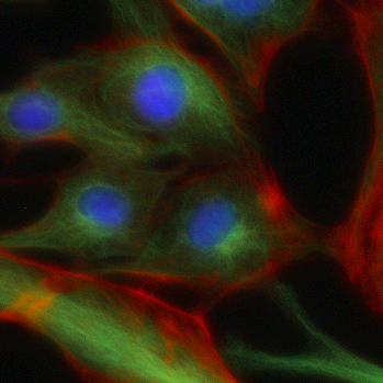 The ability to microscopically visualize the tiniest of molecules provides the backbone of basic medical research.
Michael Mancini, Ph.D., director of Baylor College of Medicine’s Integrated Microscopy Core, explains the core’s strong set of automated platforms and new technologies are revolutionizing scientific discovery and hastening research in the areas of drug development and pathology.
“We provide not only the instrumentation but also design and develop an appropriate assay to achieve project goals. We help investigators identify the best platform to support their needs and determine the most efficient way to help them get answers to their research questions,” Mancini said. “Most institutions have basic microscopy cores, but what sets us apart is a strong set of automated platforms and access to new technologies through various corporate partners.”
Read the complete interview with Mancini on VIICTR.org.
Additional Resources
What inspires biomedical research?
Cultivating a new generation of clinical investigators.
Explore the Virtually Integrated Institutions for Clinical and Translational Research.
Online resource helps connect researchers, resources
Advancing innovative research for Baylor scientists
Dr. Maria Monica Gramatges says being a physician and scientist is the best of both worlds.