Researchers combat Zika-associated fetal abnormalities using microRNA
Before SARS-CoV-2 and the COVID-19 pandemic, there was the Zika virus epidemic, lasting from 2015 to 2016. The Zika virus can cause serious birth defects and abnormalities. During the epidemic, one of the most striking results of Zika virus in pregnant women was the increase in offspring with microcephaly or a head much smaller than expected, a condition that can result in abnormal brain development.
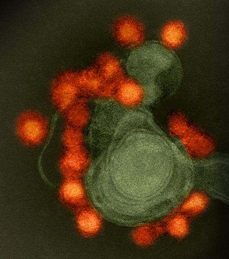
While the Zika virus epidemic has ended, future outbreaks are inevitable as most of the world’s population lives in areas where the Zika virus mosquito thrives. Researchers in the Aagaard Lab at Baylor College of Medicine studied how the virus persists in the placenta for long periods and how to mitigate it. Their findings were published in the American Journal of Obstetrics and Gynecology.

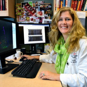
“At the maternal-fetal interface, there are immune niches and viruses that might take advantage of how long they are able to persist in the placenta,” said Dr. Enrico Barrozo, postdoctoral associate in the Department of Obstetrics and Gynecology at Baylor and first author of the paper.
Zika virus has a high maternal-fetal transmission rate leading to a number of fetal health issues, not just microcephaly. Infections can cause significant changes in early immune development. If a virus or microbe is able to persist for months, it can potentially have more of an effect on early immune development. Pregnant women infected with Zika virus have an increased chance of early pregnancy loss as well as a variety of other defects.
“The spectrum of babies isn’t just around microcephaly, but there are a number of different kinds of anomalies that can accompany Zika virus infection,” said Dr. Kjersti Aagaard, professor and maternal-fetal medicine expert in the Department of Obstetrics and Gynecology at Baylor and Texas Children’s Hospital. “They range from hearing loss to much more complicated and morbid conditions.”
Researchers used primary human placenta cells, a mouse fetal model of the disease and a new technology of spatial transcriptomics which allows researchers to zoom in on placental microenvironments or niches. This is the first robust spatial transcriptomics study using mouse placenta. The study was conducted in three phases:
First, the team used a model of primary human placenta cells cultured in the lab to look at microRNA activity in response to Zika virus infection experimentally. microRNAs are cell-produced molecules that have a variety of functions, including playing roles in viral infections.
They found that Zika virus infection of human placental cells led to an unexpected disruption of placental microRNA regulation networks.
Second, they studied Zika virus disease development in a germ-free mouse model. Mice are usually resistant to Zika virus, but in this mouse model that does not have any live microbes, they were susceptible.
Treatment of these Zika virus-infected mice with enoxacin, an FDA-approved antibiotic that enhances placental microRNA pathways, abolished placental Zika virus persistence and rescued the associated microcephaly.
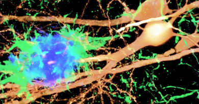
Third, they used spatial transcriptomics to detect differences in the placental immune microenvironments in the mouse model. They found niches with a significant increase of components of the immune response, such as the complement cascade and changes in TGF-β gene expression.
After treating with enoxacin, the Zika virus-associated transcriptional changes in placental immune microenvironments were no longer observed.
“It’s really important to understand how viruses are, or are not, able to set up a niche in the placenta at the maternal-fetal interface,” Barrozo said. “We were very lucky to have this unique model with Zika virus susceptibility, and that we were able to untie the antibiotic part of enoxacin from its microRNA activity.”
Moving forward, the research team hopes to uncover the parts of the microbiome that are associated with viral resistance and early immune development and when that occurs in pregnancy.
“Our next steps are developing an exciting new line of work around the animals that don’t have microbes (gnotobiotic animals) and understanding why they are susceptible to Zika virus infection,” Aagaard said.
Other contributors to this work include Dr. Maxim Seferovic; Dr. Mark Hamilton; David Moorshead; Dr. Michael Jochum; Trang Do; Dr. Derek O’Neil; and Dr. Melissa Suter.
Funding: NIH-NICHD (R01HD091731), NSF Postdoctoral Fellowship (#2208903) and a Career Development Award from the American Society of Gene & Cell Therapy.
Follow From the Labs on Twitter @BCMFromtheLabs.