Video of the Month: A beating developing mouse heart
The lab of Dr. Irina Larina develops and applies live high-resolution imaging and dynamic analysis that enable the study of the mammalian heart at early developmental stages. This image shows an application of optical coherence tomography, a highly innovative method developed in her lab, that in combination with live embryo culture protocols allows for the visualization of the entire live mouse embryo with single cell resolution, which is currently not possible with any other imaging technique.
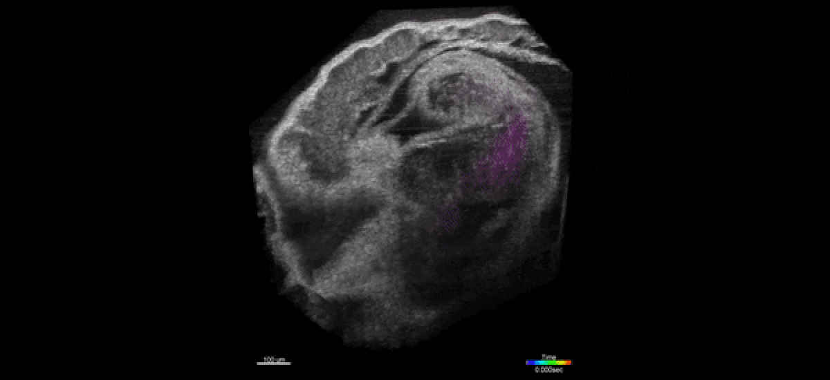
“We can visualize beating embryonic hearts volumetrically at the rate of 100 volumes per second, which provides both quantitative and qualitative information about the heart tube dynamics and allows for rapid identification of mutant embryo physiology,” Larina said. “There is a number of projects in-progress in the lab based on these methods, which are aimed at understanding mechanism of cardiogenesis and mutant phenotype analysis.”
The Larina lab is also developing an in vivo 3D imaging method for visualizing the mouse oviduct and reproductive events with micro-scale spatial resolution. These measurements are currently not possible with any other method and provide a unique opportunity to explore reproductive processes from a new dynamic perspective, adding to the development of fertility treatments and contraception. Read her work about how the egg and the embryo actually travel through the fallopian tube.
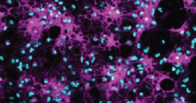
In addition, the team works in projects involving in vivo micro-scale tomography of ciliary behavior. Cilia in the oviduct are very small, between 5-10 μm in length and approximately 300 nm in diameter, and this makes their live visualization and activity in the lumen of the oviduct through tissue layers a major challenge. The Larina lab developed a functional low-coherence optical imaging technique that allows in vivo depth-resolved mapping of the cilia location and cilia beat frequency in the intact mouse oviduct with micro-scale spatial resolution.
Learn more about the research conducted in the Larina lab in her website.

Dr. Irina Larina is associate professor of integrative physiology at Baylor College of Medicine