Skip to content
Monday, June 16, 2025
Latest:
  • What is inflammation? Q&A with Dr. Mike Ren
  • A new treatment can potentially change the outlook for patients with Alagille syndrome
  • Mental health for first-time parents
  • Heart attack survivor credits Baylor doctor for a second chance at life
  • It’s a first: implementing safe and effective RSV prevention treatments significantly reduces hospitalizations in infants
Baylor College of Medicine Blog Network

  • Home
  • Momentum
    • About Momentum
    • Resources
    • Contributors
      • Taylor Barnes
      • Dana Benson
      • Molly Chiu
      • Julie Garcia
      • Graciela Gutierrez
      • Anna Kiappes
      • Andy Phifer
      • Homa Shalchi
      • Melissa Tucker
    • Contact Us
  • From The Labs
    • About From the Labs
    • Follow From The Labs on Twitter
  • Policywise
    • About PolicyWise
    • Follow @BCMEthics on Twitter
  • The Stitch
    • About The Stitch
  • Progress Notes
    • About Progress Notes
    • Progress Notes Editors
    • Connect With Us
  • bcm.edu
Still image to accompany July 2019's Video of the Month (Hyser lab) Cells expressing a fluorescent calcium sensor (green) were infected with rotavirus, a virus that causes acute gastroenteritis. The rotavirus-infected cells (pink) trigger an intercellular calcium wave that increases calcium in surrounding uninfected cells. (Scale bar = 100 μm; courtesy of the Hyser lab).
From The Labs

Video of the Month: A calcium storm

July 2, 2019 calcium indicator,  gastroenteritis,  Research,  rotavirus,  video

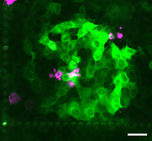
From the Labs opens July 2019 with a Video of the Month, showing the calcium ‘storm’ rotavirus triggers in infected cells. Calcium signaling in rotavirus

Read More
From The Labs

Through the looking glass, fluorescent signals reveal the workings of a fly’s brain

August 19, 2016 ASAP,  calcium indicator,  electrodes,  fluorescent voltage indicator,  Neuroscience,  two-photom microscopy

[listen to audio] Bain activity is fast. Millisecond fast. When a person, a mouse or a fly sees an object, neurons in their brains display

Read More

From the Labs: Image of the Month

Blood vessels (magenta) intertwined with metastatic medulloblastoma tumor cells (green) nestled within the protective layers that surround the mouse spinal cord. From the Labs: a closer look at metastatic medulloblastoma

Healthy Habits: A DOC-umentary Series

https://www.youtube.com/watch?v=SlNiMWHUhbc

Archives

Search

©1998 - 2023 Baylor College of Medicine®
One Baylor Plaza, Houston, Texas 77030
(713) 798-4951