These cells are critical to the endometrium’s remarkable ability to regenerate

Scientists have long known that the endometrium, the tissue lining the inside of the uterus, has a remarkable ability to regenerate. In humans, this tissue is rebuilt every month after menstruation, and in both humans and animals, it must also repair itself after pregnancy and childbirth.

“Throughout a woman’s lifetime, the endometrium has the potential to undergo about 300 to 400 cycles of growth, regeneration and shedding,” said Dr. Diana Monsiviais, associate professor of pathology and immunology at Baylor College of Medicine and corresponding author of this work. “But the actual growth factors and cells that are leading to this coordinated regeneration are not well understood. Here we investigated the role cells with high levels of an enzyme called ALDH (ALDH-positive cells) play in the endometrium’s regeneration.”
Relevance to endometriosis
Understanding these factors is essential to treat endometriosis, a condition in which the lining of the uterus grows outside its normal location, often on the ovaries, the fallopian tubes, uterine body or attached to the intestines or the lining of the abdominal cavity. This typically causes inflammation, severe pelvic pain and infertility that significantly reduce quality of life.
Endometriosis affects more than 190 million women and girls worldwide with pain that can lead them to pull away from school, work and home.
Current treatments have limited benefits and largely depend on hormonal therapies, which are partially effective at reducing pain, and may cause side effects, lead to therapy resistance and are unsuitable for some patients, particularly those trying to become pregnant. Many patients also undergo repeated surgeries because the disease often returns. There is a great need for improved treatments that cause remission of the disease, reduce pain and restore health to this underserved medical need. The current study published in eLife is a step in that direction.
Which cells help the uterus regenerate?
Monsivais and her colleagues at Baylor focused on cells in the uterus that are responsible for its periodic, normal regeneration; specifically, they investigated whether ALDH-positive cells act as stem cells in the uterus.
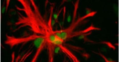
“We examined both mouse and human endometrial tissue and used several laboratory techniques to identify and track cells with high ALDH activity,” Monsivais said. “Stem cells are typically able to survive for long periods, generate many new cells and rebuild tissues after injury. We found that cells with high ALDH activity showed many of these characteristics. When grown in the laboratory, these cells were much better at forming miniature uterus-like structures called organoids than cells with low ALDH activity. They also expressed genes commonly associated with stem cells and tissue regeneration.”
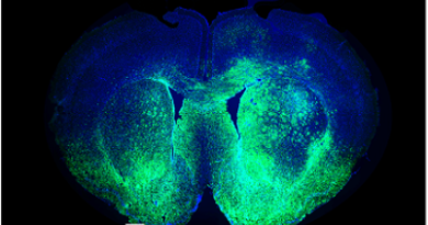
The team took a developmental approach to better understand how the endometrium fulfills its functions. The researchers discovered that cells with high ALDH activity persisted for long periods and produced new cells that contributed to both the glandular and surface layers of the endometrium. During postnatal development, ALDH-positive cells expanded as the uterus matured. In adult mice, they continued to participate in normal tissue maintenance and regeneration.
We also found that the ALDH-positive stem-cell-like population is sensitive to hormonal signals and may adjust its behavior according to the uterus’s changing needs. These findings suggested that the cells behave as long-lived progenitors or stem cells that help maintain uterine health throughout life,” Monsivais said.
Furthermore, after childbirth, when the uterus must repair tissue left behind after the placenta detaches, the team observed ALDH-positive cells in areas undergoing active repair. Their presence during healing suggests they help rebuild damaged tissue and restore normal uterine structure following delivery.
To test whether these cells truly contribute to uterine development, the researchers selectively removed ALDH-positive cells in mice. Mice lacking these cells had fewer endometrial glands and lower levels of FOXA2, a protein required for healthy gland function. In laboratory-grown organoids, eliminating ALDH-positive cells caused structural breakdown and cell death.
These results demonstrate that the cells are not just associated with tissue regeneration but also are required for normal development and maintenance of the uterine lining,” Monsivais said.
ALDH-positive cells and the human endometrium
Importantly, the team also observed these findings in human endometrial cells with high ALDH activity. These cells formed organoids more efficiently and survived longer in culture than cells with low ALDH activity. They also displayed gene activity patterns linked to stem-cell properties and tissue growth, while cells with low ALDH activity showed features of more mature, specialized cells.
This similarity between mice and humans strengthens the evidence that ALDH marks an important regenerative cell population in the endometrium.
“We determined that ALDH-positive cells in the healthy endometrium possess regenerative potential that is critical for endometrial development and function,” Monsivais said.
We are currently looking at ALDH-positive cells in the context of endometriosis and determining whether targeting these or other cells could lead to a much-needed treatment for this debilitating condition.”
Other contributors to this work include first author Suni Tang, Anna Catherine Unser, Peixin Jiang, Sydney E Parks, Genesis J Herrera, Ting Geng, Linda Alpuing Radilla, Brooke A Thigpen and Xiaoming Guan, all affiliated with Baylor College of Medicine.
The study was supported by the Eunice Kennedy Shriver National Institute of Child Health and Human Development (grants R01-HD105800, R01-HD099341), a Next Gen Pregnancy Award (NGP10125) from the Burroughs Wellcome Fund, a Lalor Foundation Postdoctoral Fellowship, a P30 Cancer Center Support Grant (NCI-CA125123), a CPRIT Core Facility Support Award (CPRIT-RP240432) and NIH grants (CA125123, OD036336, DK56338, CA125123, P30 CA125123, S10OD030414).
Follow From the Labs on X, BlueSky and Instagram!
Would you like to receive From the Labs via email?
Follow this link.