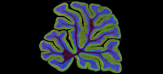
The cerebellum as a source of generalized convulsive seizures
Recurrent seizures are debilitating and sometimes fatal. While the beginning and manifestation of seizures can vary significantly, of more than 25 categories of seizure presentations within epilepsy, those with convulsions are perhaps the most disruptive and generally the most feared by patients and caregivers. Unfortunately, convulsive seizures also are the most commonly occurring type of generalized seizures.
Little is known about the source or the precise mechanism by which these seizures originate, making it difficult to develop effective treatments for these patients, but a study led by Dr. Roy Sillitoe, professor of pathology and immunology and neuroscience at Baylor College of Medicine and investigator at the Jan and Dan Duncan Neurological Research Institute (Duncan NRI) at Texas Children’s Hospital, provides new insights into how seizures happen. The study was published in Communications Biology.
To our knowledge, this is the first study to implicate the cerebellum in altering the activity of a specific group of neurons in a brain area called the ventral posteromedial nucleus (VPM) in the thalamus, which then initiate generalized convulsive features,” Sillitoe said.
“These findings not only deepen our understanding of how seizures originate but are expected to have a wider impact on several neurological diseases. Moreover, this study opens the tantalizing possibility of targeting cerebellar circuits to treat generalized epilepsies and other neurological disorders.”
A reciprocal cortico-thalamic loop is central to generalized convulsive seizures
While each brain region performs discrete functions, they do not act in isolation. In fact, many regions are in constant communication with one another through a multitude of interconnected neural networks. The reciprocal cortico-thalamic loop is part of one such network that links neurons in the forebrain – namely the cerebral cortex – which controls cognition, memory, language and emotion processing with the thalamus, an-egg-shaped midbrain structure that acts as a relay center for motor and sensory information.
This cortico-thalamic neural circuit is important for several critical brain functions such as sleep and learning and plays an unequivocal role in different disease states (for example, certain types of seizures and tremors).
Thalamic neurons, a critical part of this circuit, are the focus point of seizure activity. However, exactly how these thalamic neurons generate or propagate generalized seizures was unclear.
Facial twitching is often among the first manifestations in generalized convulsive seizures in patients; therefore, the authors chose to study the VPM thalamic circuits, which are tightly interconnected to another part of the brain called the facial somatosensory cortex and have been previously suspected to play an important role in various types of epilepsies.
The majority of thalamic VPM neurons are active and contribute to seizures
Using optogenetics – a technique that applies light to control genetically manipulated neurons – and drug injection techniques, the researchers found that neuronal activation specifically in the VPM, but not in other surrounding thalamic nuclei, caused severe convulsive seizures in mice.
Studies in animal models have established that a proportion of cells within the epileptic brain remain largely normal during seizure episodes. Here, the team found that a higher proportion – as many as 80% – of VPM neurons may change their activity and actively contribute to initiating or sustaining seizures. Within this population, the activity of the affected neurons can take on different forms, altogether suggesting a high level of heterogeneity in neural responses as well as the identity of neurons that respond versus those that remain unchanged.
The cerebellum is a major source of stimulation for VPM neurons during seizures
To their greater surprise, the team found that cerebellar neurons showed the greatest ability to drive seizures among the possible contributing brain regions. Viral tracing experiments revealed specific signals that traveled from the cerebral cortex and cerebellar regions to VPM neurons that were activated during seizures. Interestingly, when they inhibited the activity of the cerebellar output pathway with lidocaine, VPM neurons stopped inducing seizures.

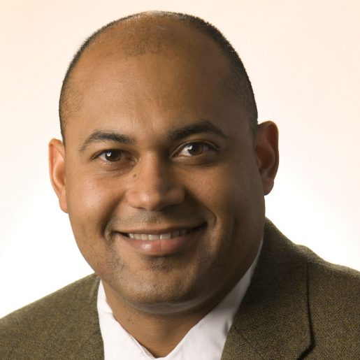
“Identifying a new critical neural circuit from the cerebellum to the VPM neurons in the thalamus as the source of seizures is indeed exciting. It provides fundamental insights into how major brain nodes are interconnected and regulated for optimal function and behavior,” said first author Dr. Jaclyn Beckinghausen, who worked in the Sillitoe lab while developing this project.
“It is becoming increasingly apparent that the cerebellum, Latin for ‘little brain,’ may not be so little in its ability to drive distinct behaviors under normal and diseased states,” Sillitoe said. “Given its increasingly diverse and complex roles in motor and non-motor functions and, accordingly, its contributions to diseases ranging from ataxia, dystonia and tremor to autism, schizophrenia and seizures, one wonders what other big mysteries this powerful ‘little brain’ might be hiding.”
Others involved in the study were Joshua Ortiz-Guzman, Tao Lin, Benjamin Bachman, Luis E. Salazar Leon, Yu Liu, Detlef H. Heck and Benjamin R. Arenkiel. They are affiliated with one or more of the following institutions: Baylor College of Medicine, Texas Children’s Hospital and University of Minnesota Medical School.
This work was supported by funds received from Baylor College of Medicine, Texas Children’s Hospital, the Hamill Foundation and grants from the National Institutes of Neurological Disorders and Stroke, National Institute of Mental Health, the Dystonia Medical Research Foundation and by the Eunice Kennedy Shriver National Institute of Child Health & Human Development of the National Institutes of Health for the use of the Cell and Tissue Pathogenesis Core and the Neuroconnectivity Core.
Disclosure: Dr. Sillitoe serves on the Interim Governing Board of the Raynor Cerebellum Foundation, Fort Worth, Texas.
Read more here.