When it comes to testing for contaminants in our environment, the more rapid and sensitive the test, the better. That is why researchers at Baylor College of Medicine have created new cell line models allowing fast, sensitive, direct observations of what happens when estrogen receptors are exposed to estrogen-disrupting chemicals.
Estrogen-disrupting chemicals can be found in water, soil or even food. Image credit: Pixabay.
Estrogen-disrupting chemicals (EDCs) are substances found in our environmental exposures to water, soil or even food. These types of chemicals are known to interfere with the endocrine system, which is responsible for many bodily functions, including growth, development and metabolism. Additionally, exposure to these chemicals can lead to certain illnesses, including some forms of cancer.
“Our new cell models allow us to detect a wider range of these types of chemicals,” said Dr. Adam Szafran, an instructor in molecular and cellular biology at Baylor. “Improving on past cell lines, we were able to maintain the same endpoints as the original assay, allowing for the direct observation of multiple mechanistic steps of estrogen receptor signaling when exposed to chemical mixtures.”
The findings, appearing in iScience, describe how researchers tweaked the cell lines that have been used for similar testing in the past so that the level of estrogen receptor expression can be controlled.
While the ability to detect strong and moderately strong estrogens remains unchanged compared to previous cell lines, the new cell lines detect up to 90% of even very weak estrogen-disrupting chemicals, markedly improving the detection rate of previous assays.
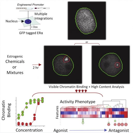
Testing estrogenic responses has long been conducted first in living mouse models, a very slow and expensive process. While quicker and less expensive, early in vitro –in a test tube in the lab – assays answered only one question, whether estrogenic activity was detected. More sensitive cell lines were produced to replace that type of testing, and in the current study, Szafran and colleagues have improved the assays to express either inducible C-terminal or N-terminal (parts of proteins having differing chemical properties) GFP-tagged estrogen receptors, allowing for direct visualization of activity when exposed to estrogen receptor hormones, ligands or endocrine disrupting chemicals.
Graphical abstract. Image courtesy of the authors/iScience, 2022.
“We are exposing the cell lines to a mixture of well-known toxins commonly used in testing by the Environmental Protection Agency. The data points are complex and robust and required numerous cell line-based assays that take hours or weeks to collect and process data. But now, our new models can make the call on potential estrogen-disrupting chemicals ‘hits’ within in minutes and simultaneously report on potential mechanisms of action data,” said Dr. Michael Mancini, professor of molecular and cellular biology and director of the Integrated Microscopy Core at Baylor.
We are looking for a wide range of responses from these assays, determining whether test samples are activating estrogen receptors (agonists) or stopping receptors (antagonists) from producing responses,” Mancini said.
In a boost to efficiency, the published work shows how these dueling questions can be answered in a single experiment due to the biology captured by the updated cell lines.
Dr. Michael Mancini
Moving forward, Mancini said the goal is to utilize models to first characterize the estrogenic activity of certain chemicals or mixtures before they are put into the environment.
“In addition, we can test environmental samples. These new cell lines now allow us more sensitivity to detect moderate or weak chemicals and to do so while keeping the approach a high-throughput assay,” he said.
Furthermore, with the arrival of new NIH-funded, very-high-throughput/versatile confocal and robotic equipment in-house, these types of sensitive assays are now possible for a wide range of projects from many labs.
Other contributors to the study include Dr. Fabio Stossi, associate professor of molecular and cellular biology, and Maureen Mancini, research associate in molecular and cellular biology, both with Baylor.
Funding sources include NIEHS (Project 4 and Superfund Research Program grant, P42ES027704), CPRIT-funded GCC Center for Advanced Microscopy and Image Informatics (RP170719), CPRIT (RP150578), and support from the Center for Precision Environmental Health (P30ES030285). Imaging was also supported by the Integrated Microscopy Core at Baylor College of Medicine with funding from NIH (DK56338 and CA125123) and the Dan L Duncan Comprehensive Cancer Center.