What patients need to know about skin grafts
Using skin grafts for wound coverage is a foundational technique in plastic surgery. The history of skin grafting traces back thousands of years. In fact, grafting techniques are well documented in ancient Egypt and India. The 19th and 20th centuries saw rapid advancement and standardization of grafting techniques. Today, plastic surgeons rely on various types of natural and synesthetic skin, donor sites and preparation methods to care for patients with burns, wounds, non-healing ulcers and skin defects that require coverage with grafting.
Types of skin grafts
 The skin is composed of three layers: the outer layer epidermis, the middle dermis and the deep hypodermis. When discussing grafting, surgeons typically divide grafts into two main categories: split-thickness and full-thickness. Split-thickness skin grafts are grafts involving the epidermis and a portion of the dermis. Full-thickness skin grafts involve the epidermis as well as the entire dermis.
The size and depth of the wound, location on the body and aesthetic match all play into a surgeon’s decision when selecting which type of skin graft to use for a patient. Typically, split-thickness skin grafts are used to cover large areas of the body (i.e., trunk or extremities) that do not involve the joints. In contrast, full-thickness skin grafts are reserved for cosmetically sensitive sites (i.e., face) and areas that involve the joints. For example, a split-thickness skin graft would be used to reconstruct the skin of a patient with a large wound on the leg, whereas a full-thickness skin graft would be used to reconstruct the tip of a patient’s nose after a skin cancer excision. Importantly, split-thickness skin grafts usually experience more change over time, and thus are better suited for large areas that can sustain skin shrinkage. They also have a higher rate of healing in the general population as compared to full-thickness skin grafts, and are more fragile to trauma due to the thick layer of dermis within the graft.
Surgeons usually select grafts from specific areas of the body depending on the type of graft that is needed. Common donor sites for split-thickness grafts include the anterior and lateral thigh. The skin around the ear and clavicle, the upper eyelid and the groin are all areas used to harvest full thickness skin grafts. Surgeons usually use a small machine called a Dermatome to obtain split-thickness grafts during surgery when a patient is under general anesthesia. As full-thickness skin grafts are usually used to cover small areas, surgeons can remove and harvest this skin when in clinic under local anesthesia. Usually, donor sites for full-thickness skin grafts are closed primarily with stitches, whereas split-thickness grafts are left open and can close on their own with time.
What makes a skin graft successful?
The survival or “take” of both split-thickness skin grafts and full-thickness skin grafts is dependent on the ingrowth of capillaries from the recipient wound bed and the development of new blood vessels. This process is traditionally defined in three stages:
- The first stage is known as imbibition, which takes place during the first 48 hours. During this phase, the graft absorbs moisture and fibrinogen from the recipient bed, which serves to adhere the graft to the wound.
- The second is inosculation, which is an ill-defined phase where capillaries begin to extend into the graft.
- The third is revascularization, which begins after 72 hours. At this point, a combination of new blood vessel formation and connection of capillaries from the donor site to the recipient skin begin to bring blood flow to the graft. Cellular turnover, swelling and color change are all common at this point as the graft begins to stabilize and be incorporated into the surrounding tissue.
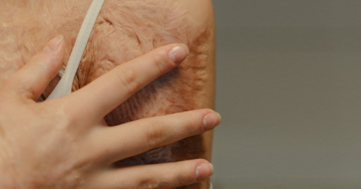
Taking care of skin grafts and donor sites
While every surgeon differs on healing protocols, most agree that grafts should not be left open to air immediately following surgery. Moist and occlusive dressings, including bolsters, wound vacs and petrolatum-infused dressings, are standard after graft placement. Alternatively, donor sites for STSGs are usually covered for around a week before being exposed to air. Common reasons for graft failure include mechanical forces, such as shearing movement, or issues related to revascularization such as fluid collection. Patient conditions such as diabetes or peripheral vascular disease can also impact a graft’s survival, as these diseases interfere with blood supply. It is important to always follow the surgeon’s guidance after receiving a skin graft.
While surgeons have traditionally relied on autografts, or skin taken from a person’s own body, new developments have led to the rise of skin substitutes from human donors, animal donors and synthetic material. These new materials include skin equivalents, wound matrixes, tissue scaffolding devices and biologic dressings. These materials can be used in a single or dual layer to replicate the epidermis or dermis or can be used to mature a recipient wound bed to help improve the take of a graft.
Whether full- or split-thickness, autologous or synthetic, skin grafting will continue to be important in plastic surgery to achieve effective healing and recovery for patients in need.
Learn more about Baylor Medicine Plastic Surgery.
By Katie Cardenas, PA-C, an instructor in the Division of Adult Plastic Surgery at Baylor College of Medicine.