The writhing and coiling of DNA drives cell activity
By Ruth SoRelle, M.P.H.
Using a multidisciplinary approach, researchers, led by those at Baylor College of Medicine, revealed in unprecedented detail the three-dimensional structure of biologically active DNA. A report on their work appears online in the journal Nature Communications.
“The beautiful double-helical structure we all know and love is not the actual active form of DNA,” said Dr. Lynn Zechiedrich, professor in the department of molecular virology and microbiology, and co-contributing author with Dr. Wah Chiu, professor in the Verna and Marrs McLean Department of Biochemistry and Molecular Biology.


Chiu and Zechiedrich, collaborating with Dr.Steven Ludtke and Dr. Michael Schmid, also of Baylor College of Medicine, and Dr. Sarah A. Harris of the University of Leeds in the United Kingdom, examined tiny DNA minicircles (now known as “MiniVectors,” a named coined by the team and commercially available) containing only 336 base pairs, using methods from chemistry, physics, math and computer modeling. Base pairs are the building blocks of genetic material.
Previous studies on short fragments
“Previous studies were on short fragments (6 – 12 base pairs) of linear DNA, but human DNA is constantly moving around in your body – and it coils and uncoils. You can’t coil linear DNA and study it, so we had to make circles so the ends would trap the different degrees of winding,” said Zechiedrich.

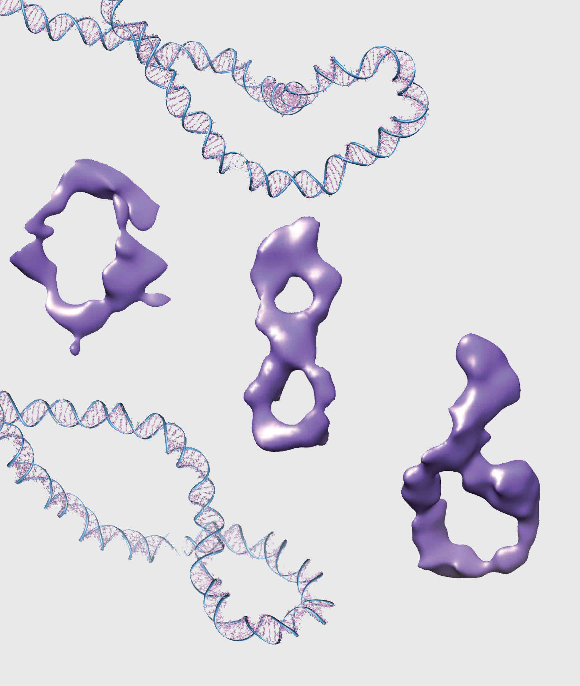
Each cell in the human body holds about a meter of DNA (ten million times longer than the tiny circles the team made). In their study, they wound or unwound a single turn at a time the DNA double helix comprising their circles and asked how the winding changed what the circles looked like, using very powerful microscopes.

Tiny, twisted DNA circles
The researchers devised a test to make sure that the tiny twisted up DNA circles that they made in the lab were biologically active. They used purified human topoisomerase II alpha, an essential enzyme that manipulates DNA and important target of anticancer drugs.
This enzyme relieved the winding stress from all of the supercoiled minicircles, even the most coiled ones, which is its normal job in the human body. This result means that the circles must look and act like the much longer DNA that topoisomerases encounter in human cells.

“These enzymes don’t do anything to linear DNA because it’s not coiled up,” said co-author Dr. Daniel J. Catanese, Jr., also of Baylor.
Cryo-electron tomography
Dr. Rossitza N. Irobalieva, the co-lead author on the publication, who is also at Baylor, used cryo-electron tomography, a powerful microscopy technique that involves freezing biologically active material, to provide the first three-dimensional structures of individual DNA molecules. She saw that the coiling caused many different shapes.
“Some of the circles had sharp bends, some were figure 8s, and others looked like racquets or sewing needles. Some looked like rods because they were so coiled,” said Irobalieva.
“Being able to observe individual DNA circles allows us to understand the different structures of biologically active DNA. Each of these different structures facilitates how DNA interacts with proteins, other DNA and RNA, and anticancer drugs, adapting to the cell processes required,” said Dr.Jonathan Fogg, the other lead author of the publication, also of Baylor College of Medicine.

While the researchers expected to see the opening of base pairs when the DNA was underwound, they were surprised to see this opening for the overwound DNA. They hypothesized that this disruption of base pairs may cause flexible hinges, allowing the DNA to sharply bend, perhaps helping to explain how a meter of DNA can be jammed into a single human cell.
“The next step is to start adding the other components of the cell or anticancer drugs to see how the DNA shapes change,” said Fogg.
Other contributors include Muyuan Chen, Anna K. Barker, all of Baylor College of Medicine, and Dr. Thana Sutthibutpong, of the University of Leeds.
Funding for this work came from the National Institutes of Health (Grants P41GM103832 and P50GM1003297 to W.C.; Grant R01GM080139 to S.L.; and Grants RO1A1054830 and R56AI054830 to L.Z.), the Human Frontier Science Program to L.Z., Biotechnology and Biological Sciences Research Council grant (BB/I019472/1) to S.A.H., and an NIH training grant (5T90DK070121-05) through the Gulf Coast Consortia to R.N.I. The MiniVectors were made available by Twister Biotech, which now produces them commercially.