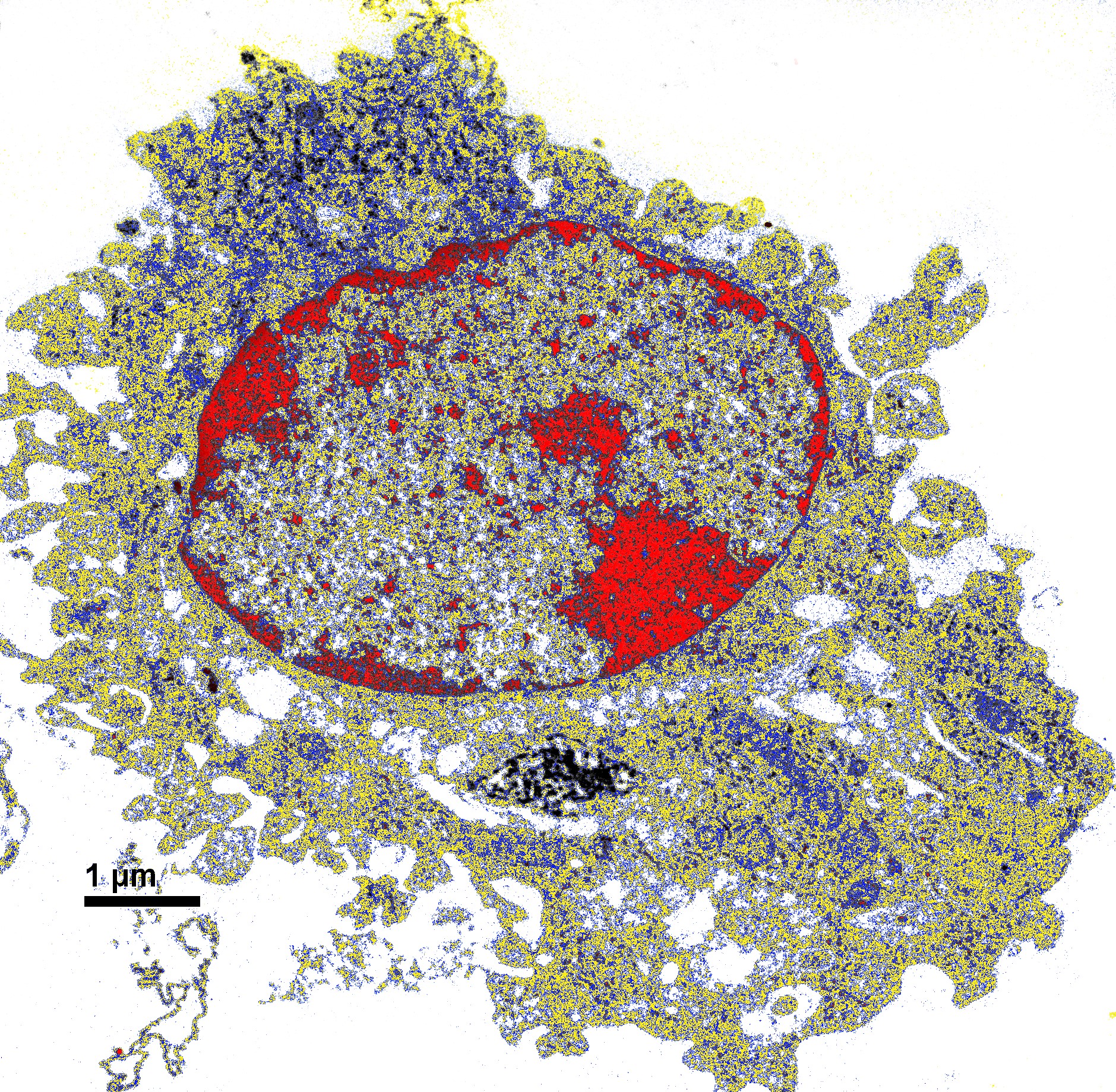
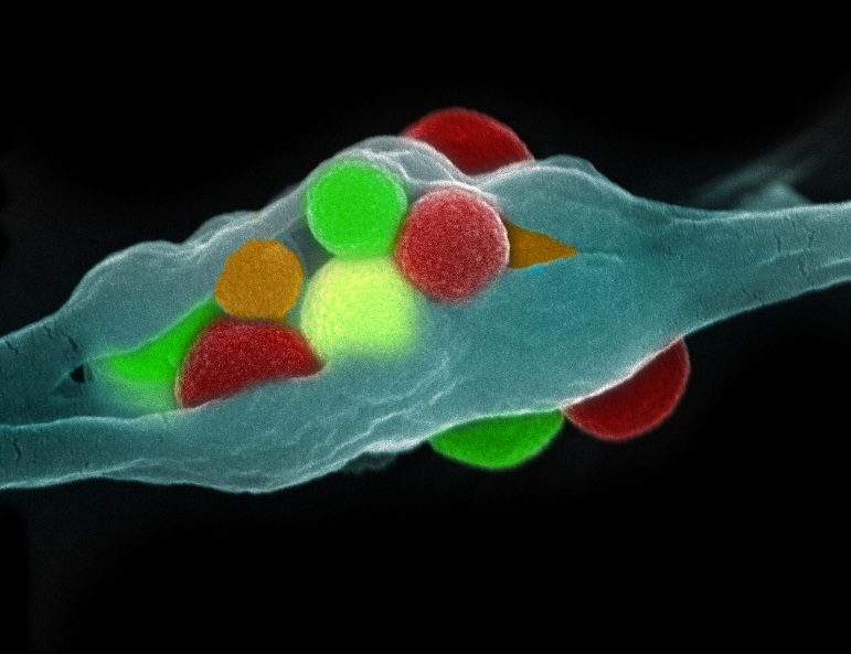
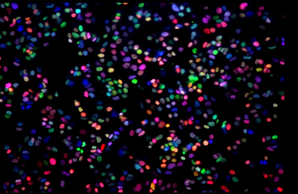
Image of the Month: Mouse breast tumor cells
Mouse mammary carcinoma 4T1 cells in pseudo-colored scanning electron micrographs. These mouse breast tumor cells (shown in these pseudo-colored scanning electron micrograph, are used to
Read More
Mouse mammary carcinoma 4T1 cells in pseudo-colored scanning electron micrographs. These mouse breast tumor cells (shown in these pseudo-colored scanning electron micrograph, are used to
Read MoreA multi-institutional team spanning Baylor College of Medicine, Rice University, Stanford University, and the Broad Institute of MIT and Harvard has reported the first successful
Read MoreBy Ruth SoRelle, M.P.H. Autopsies on smokers who died with severe emphysema reveal lungs that are black in appearance. These emphysematous lungs strain breathing structures
Read MoreCell-based trafficking of nanoparticles is achieved through in situ uptake and transport of nanoparticles by myeloid cells. Shown here is the cell membrane of a
Read MoreDNA unwinds from Chromosome 20 at the 20q13 locus. SALL4 plays important roles in hematopoiesis (formation of blood cells) and leukemogenesis (initiating the blood cancer
Read MoreMacrophages, gatekeepers of tissue integrity, are able to communicate at a distance through cellular connections coined Tunneling NanoTubes, membrane channels that contain actin and microtubules
Read MoreThis is an image showing a heart-shaped aggregate of microparticles, which were formulated for the development of a vaccine against Chagas disease at the Sabin
Read MoreThis image courtesy of Scott Holmes, Department of Surgery at Baylor College of Medicine.
Read MoreThe large surface area of graphene, a thin, tightly packed layer of carbon atoms, makes it an ideal scaffold to which you can attach a
Read MoreEditor’s note: Much of the research that ends up benefiting humans starts in laboratories that focus on model animals such as fruit flies. They are
Read More