Image of the Month: Human intestinal organoid
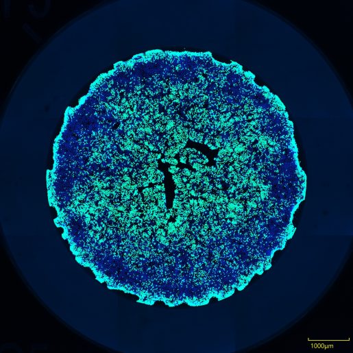
Human intestinal organoids (HIOs), also called ‘mini-guts,’ provide a laboratory model to study human intestinal epithelial biology and the processes leading to intestinal disease.
HIOs are valuable models because they retain many characteristics of the organ from which they come from, including the genetic signatures of the donor, intestinal disease and intestinal segment-specific characteristics.
HIOs have been used for screening therapeutics, studying the microbiome and its interactions with the epithelium, modeling intestinal diseases such as colorectal cancer and bacterial and viral infections, and more recently, for analysis of organoid physical characteristics in high-throughput applications.


The image this month, which was taken by Faith Sawyer, graduate student in Dr. Sarah Blutt‘s lab, is part of a recent paper published in PLOS ONE by a Baylor College of Medicine team led by the Blutt lab.
The paper describes and automated pipeline for rapidly imaging and quantifying fluorescent labeling in HIOs using a high-throughput confocal microscope and image analysis software.
This platform offers a novel approach to efficiently and rapidly image and quantify fluorescent staining and immunolabeling in HIOs and has many potential applications, including drug screening, toxicity testing, intestinal barrier integrity and transport studies, microbiome and host-pathogen interaction studies, and lineage tracking.
Follow From the Labs on X, BlueSky and Instagram!