Image of the Month: Human natural killer cells preparing to deliver a lethal strike to diseased cells
This mosaic montage of still images of live natural killer (NK) cells from the human immune system shows the cells have a skeleton of microtubules seen here in green fluorescent color. NK cells, the first responders to viral infections, use this cytoskeleton to mobilize and deliver the toxic content of lytic vesicles (here colored in red) into cells either infected by viruses or transformed during cancer progression, in order to destroy them.
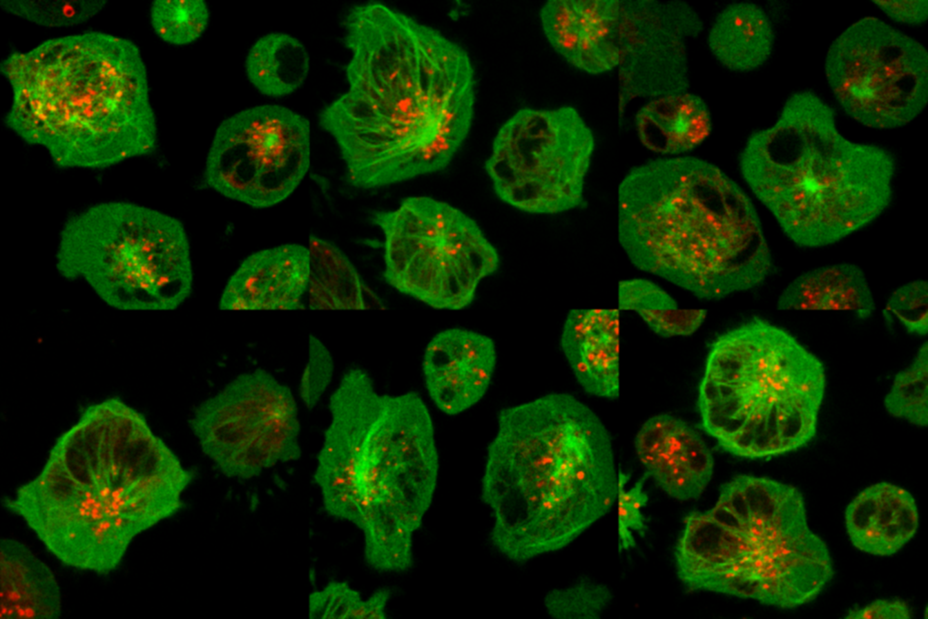
The cells have been engineered to express a green fluorescent version of the protein alpha-tubulin, the main component of microtubules inside the cells, and imaged with confocal microscopy.

Dr. Jordan Orange’s lab focuses on studying primary immunodeficiency diseases, the genetics of immunologic disease as well as the immunobiology of human NK cells. Dr. Orange’s personal clinical interests also include primary immunodeficiency diseases and NK cell deficiencies.
Dr. Orange is a professor and section head for immunology, allergy and rheumatology in pediatrics at Baylor College of Medicine and director of the Center for Human Immunobiology at Texas Children’s Hospital.
The image of NK cells was generated by Alexandre Carisey and Papiya Sinha – Orange laboratory.