Combating triple-negative breast cancer on two fronts delivers promising results
Triple-negative breast cancer (TNBC) is an aggressive subtype of breast cancer with an overall poorer prognosis than other breast cancer subtypes despite significant therapeutic advances. At Baylor College of Medicine, the labs of Dr. Jeffrey M. Rosen, Dr. Xiang H.F. Zhang and their colleagues are looking to improve treatment outcomes by better understanding the factors that contribute to the progress and the elimination of this devastating disease.
Their research work seems to have paid off. In the current study, they assessed the effectiveness of a new therapeutic approach that involved combating the cancer on two fronts simultaneously and found promising results.
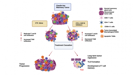
“We had previously shown in animal models that although treating the tumors with the chemotherapy drug cyclophosphamide eliminated the cancer initially, the disease came back eventually,” said Rosen, co-corresponding author of the work and Distinguished Service Professor of molecular and cellular biology and in the Dan L Duncan Comprehensive Cancer Center at Baylor. “A closer look into the tumor microenvironment revealed that the tumors that returned typically had many macrophages.”
In this study, the team explored in animal models the possibility that eliminating both the tumor cells and the macrophages would improve the chances of eliminating the tumor for a longer period of time.
“We assessed the effectiveness of a combination therapy that attacked the tumor on two fronts. On one side, cyclophosphamide eliminated tumor cells, while on the other front, another drug inhibited tumor-associated macrophages, which block the body’s immune response against the tumor,” said Swarnima Singh, first author of the paper and a student in Baylor’s graduate school of biomedical sciences working jointly in the Rosen and Zhang laboratories. “We eliminated macrophages with either a small molecule inhibitor or a monoclonal antibody toward CSF1R, a marker on these cells.”

Promising results
For several months, the team treated the tumors with the combination of drugs. Then, they stopped treatment and followed up tumor progression in the treated animals.
“We were very encouraged by the results,” Singh said. “Typically, a month after stopping single-drug treatment, the tumors returned. We were excited to see that the tumors did not return a month after stopping the combination therapy. We observed a durable response in two animal models of TNBC.”
Interestingly, the animals with a durable response also were able to prevent new tumors from growing. The researchers challenged these animals with fresh tumor cells, and 40% of them rejected the new cancer cells. The tumors did not grow. This suggested that the animals had immune memory capable of fighting back new tumor growth. In addition to primary tumors, the researchers treated lung tumor metastasis with combination therapy and found that it also eliminated cancer growth effectively.
Changing the tumor microenvironment to favor tumor elimination
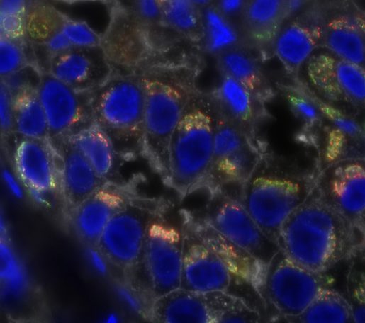
The team also investigated how the tumor microenvironment had changed in the animals with a durable response.
“We found that when we treated the tumors with one of the two drugs, which did not produce a durable response, immune B and T cells involved in fighting the tumor did not enter it, they remained in the periphery of the tumor mass,” said co-corresponding author Dr. Xiang H.F. Zhang, interim director of the Lester and Sue Smith Breast Center and William T. Butler, M.D., Endowed Chair for Distinguished Faculty. Zhang also is professor of molecular and cellular biology, a member of the Dan L Duncan Comprehensive Cancer Center and a McNair Scholar at Baylor.
On the other hand, after combination therapy, both B and T cells entered the tumor mass and remained close to each other, suggesting they were interacting to mediate tumor elimination in a durable manner.
Clinical implications
Looking to translate these findings into the clinic, the researchers investigated whether the macrophage signature they had observed in the animals models also was present in TNBC from patients.
Indeed, tumors from patients with the worse prognosis also presented high numbers of macrophages,” Rosen said. “We think this signature can help us identify patients who might be candidates for combination therapy.”
The researchers are planning to conduct a clinical trial to assess the value of their approach in treating human TNBC.
Read the full report in the journal Cancer Research.
Other contributors to this work include Nigel Lee, Diego A. Pedroza, Igor Bado, Licheng Zhang, Sergio Aguirre, Clark Hamor, Yichao Shen, Yitian Xu, Jingyuan Hu, Yang Gao, Na Zhao, Shu-Hsia Chen, Ying-Wooi Wan, Zhandong Liu, Jeffrey T. Chang, Daniel Hollern and Charles M. Perou. The authors are affiliated with one or more of the following institutions: Baylor College of Medicine, Dan L Duncan Comprehensive Cancer Center, Jan and Dan Duncan Neurological Research Institute at Texas Children’s Hospital, the University of Texas Health Science Center at Houston, University of North Carolina, Houston Methodist Research Institute and the Salk Cancer Center.
These studies were supported by grants NCI-CA148761, Breast Cancer Research Foundation, NCI CA151293, U.S. Department of Defense DAMD W81XWH-16-1-0073 and W81XWH-18-1-0574, Susan G. Komen CCR14298445 and McNair Medical Institute. Support also was provided by CPRIT grants RP160283 and RP170668, the Dan L Duncan Comprehensive Cancer Center (NCI: P30:CA125123) and Pathology Core of the Lester and Sue Smith Breast Center and T32CA203690-01A1. This project also was supported by the Advanced Technology Cores at BCM: Genomic and RNA Profiling Core (NCI: CA125123), Single Cell Genomics Core, Cytometry and Cell Sorting Core (CPRIT-RP180672), and Integrated Microscopy Core (NIH: DK56338 and CA125123; CPRIT: RP150578 and RP170719).