Discovery of CTLA-4 in dendritic cells opens new possibilities to fight cancer
By Ana María Rodríguez, Ph.D.
In the battle against cancer, T cells are the ‘foot soldiers’ and dendritic cells are ‘the generals.’ Cancer cells are not passive players in these encounters, they can fight the foot soldiers back by pushing a brake on the T cells that will turn them off. This ‘brake’ is a molecule on the surface of T cells called CTLA-4. Until now, most scientists agreed that CTLA-4 was only present on T cells and other cells of the same lineage. But Baylor College of Medicine researchers have discovered that CTLA-4 is also produced and secreted by dendritic cells. The results appear in Stem Cells and Development.

“These results are relevant to the battle against cancer because we showed that dendritic cell CTLA-4 performs a very critical regulatory function. Its presence inhibits the generation of downstream anticancer responses, whereas its absence permits robust priming of such responses. These new data provide a strong rationale to use the drug ipilimumab in new and better ways, for instance in conjunction with cancer vaccines,” said Dr. William K. Decker, assistant professor of pathology and immunology at Baylor and the Center for Cell and Gene Therapy, a member of the Dan L Duncan Comprehensive Cancer Center, and senior author of this paper.
Ipilimumab is a Food and Drug Administration-approved drug to treat melanoma. Scientists think that ipilimumab helps the body fight cancer cells by removing the ‘brake’ cancer cells place on the T cells. Ipilimumab binds to CTLA-4 on T cells, blocking signals that turn off the T cells. As a result, scientists think, T cells resume their fight against the cancer.
In this study, Baylor researchers have contributed a new piece to the puzzle of how the immune system regulates T-cell responses.
Decker and his colleagues provide solid evidence that dendritic cells, the ‘generals’ that direct the activity of the T cells, produce and release CTLA-4, which until now has been controversial.
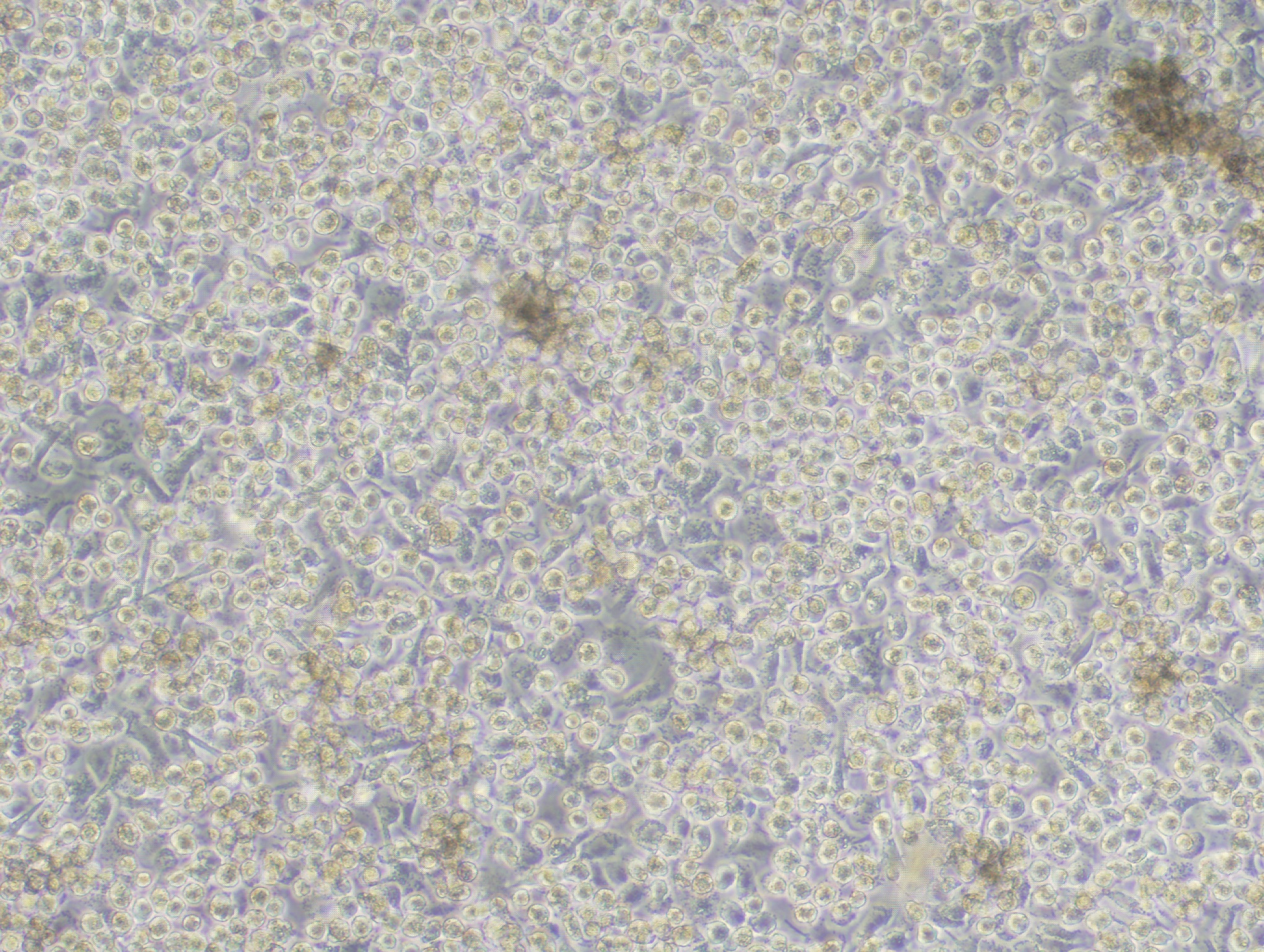
When activated, dendritic cells secrete CTLA-4-studded microvesicles into their environment. The microvesicles can bind to other dendritic cells, be internalized, and turn off the dendritic cells, which then cannot proceed to activate T cells.
CTLA-4, dendritic cells, and the anti-tumor response

“To show the relevance of turning off the dendritic cells in the body’s response against tumors, we studied a mouse model of melanoma,” said Dr. Matthew M. Halpert, an instructor in immunology and first author of the paper.
“We tested two types of dendritic cells: normal dendritic cells expressing CTLA-4 and dendritic cells treated with CTLA-4 siRNA, a strategy that dramatically diminishes the production of CTLA-4. One group of mice received melanoma cells and a vaccine against the tumor made with normal dendritic cells. A second group of mice received melanoma cells and a vaccine made with dendritic cells that produce little amounts of CTLA-4. The mice that received normal dendritic cells, which produce CTLA-4, were not able to slow down the growth of the tumor. On the other hand, the mice treated with dendritic cells that produce little CTLA-4 were able to develop an immune response that markedly limited tumor growth.”
“These results suggest that priming an immune response against melanoma in the absence of CTLA-4 triggers a response that can control tumor growth in this mouse model.”
These results have encouraged the Baylor researchers to suggest that strategies that combine taking away CTLA-4, or blocking it with ipilimumab, with specific tumor vaccines, of which many already exist in experimental settings, may result in better immune responses that can control tumor growth.
Vanaja Konduri, Dan Liang, Yunyu Chen, Silke Paust, Jon Levitt, all from Baylor, also contributed to this work. James Wing, an assistant professor at the Immunology Frontier Research Institute at the University of Osaka in Japan, was a key collaborator as well.
This project was supported by funding from Alex’s Lemonade Stand Childhood Cancer Foundation and by the Cytometry and Cell Sorting Core at Baylor College of Medicine with funding from the NIH (AI036211, CA125123, and RR024574).
Decker and Halpert own shares of Diakonos Research, Ltd.