Finding a way to grow norovirus in the laboratory
By Ruth SoRelle, M.P.H.
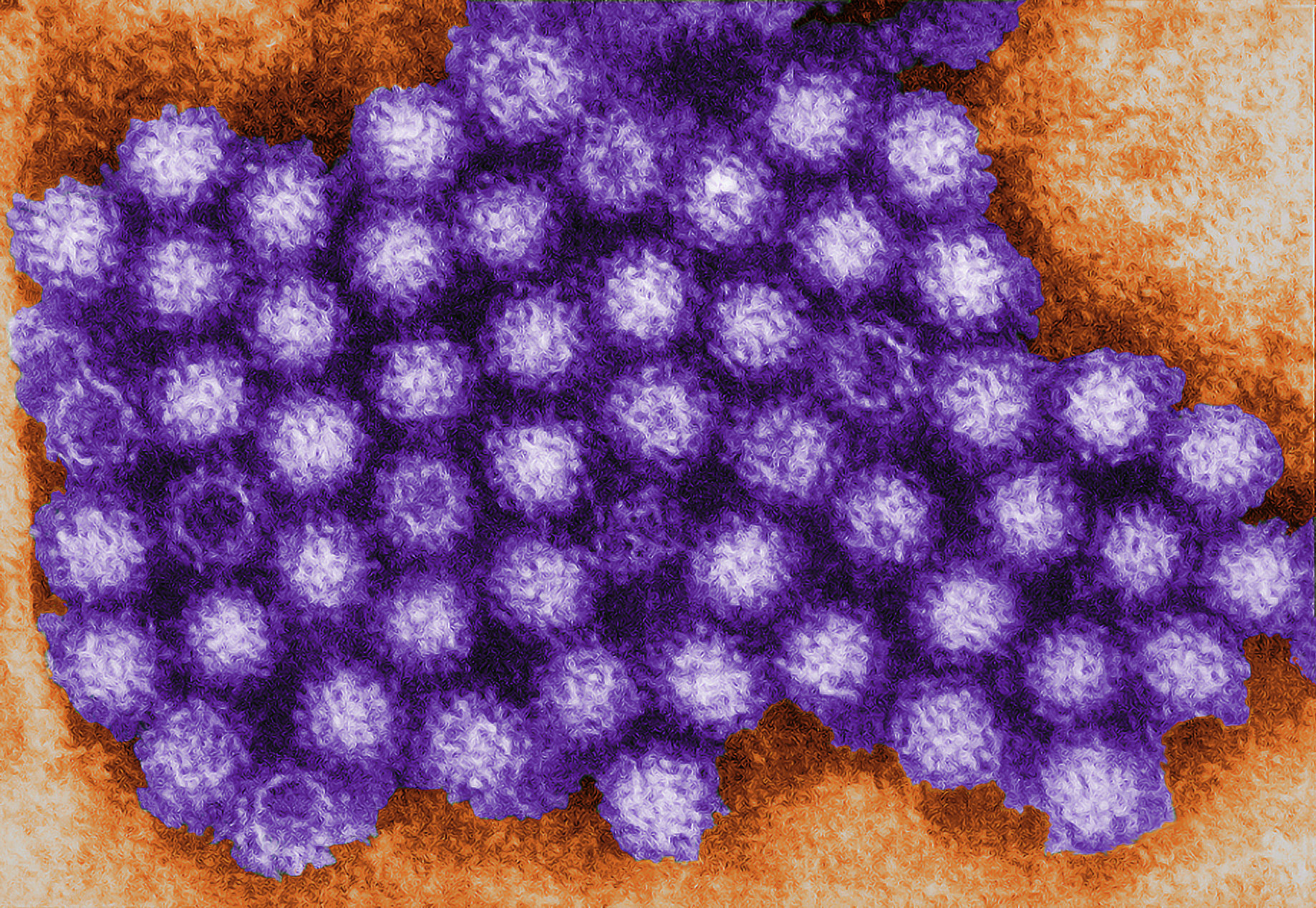
Experts seeking ways to prevent and treat infections with the human norovirus – the leading cause of gastrointestinal illness in the world – have faced a major hurdle because they cannot grow the virus in the laboratory.
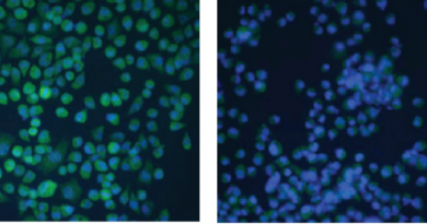
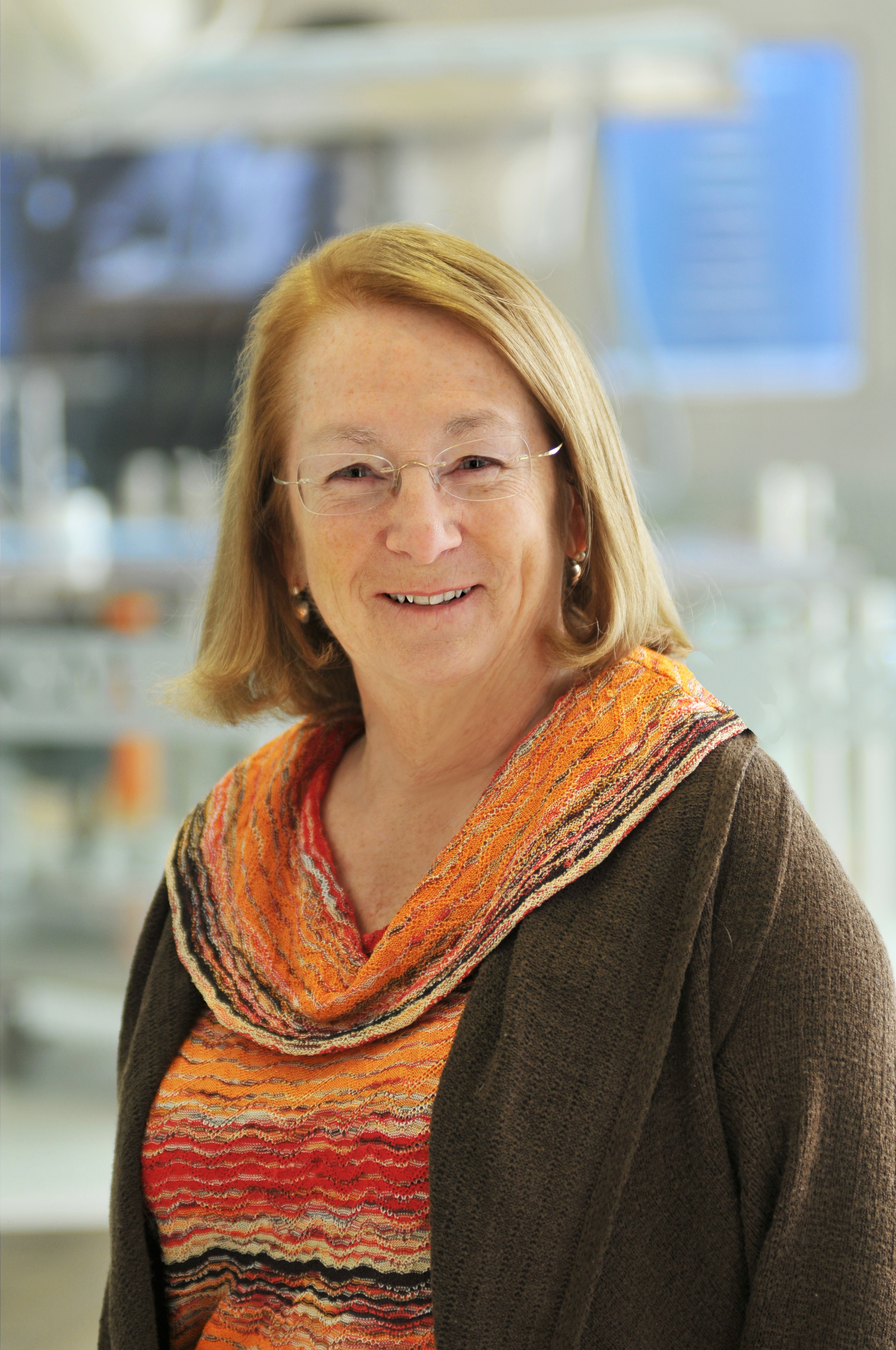
In a report that appears in the Proceedings of the National Academy of Sciences, Dr. Mary Estes, professor of molecular virology and microbiology at Baylor College of Medicine, and her colleagues describe a newly developed system using reverse genetics that allows the virus to grow in cells. This system uses a novel promoter that leads to the production of viral RNA (the viral genetic material) from DNA and supports replication of the norovirus genome and formation of virus particles, including particles that contain a green fluorescent protein. This new system will allow discovery of the viral genes responsible for human norovirus infection and disease.
Step forward
“This is a big step forward,” said Estes. “We can use this system to see if anti-viral drugs that we and other people are making work. It will also help us understand why we cannot grow the virus directly from clinical samples in culture.”
With this new system, the scientists can produce as much virus as they want.
Infectious
“We think it’s infectious,” said Estes. Building on work he began as a postdoctoral fellow at Baylor, Dr. Kazuhiko Katayama, director of the department of virology of the Institute of Infectious Diseases in Tokyo, and his colleagues have shown that a mouse form of the virus is infectious. Estes and her colleagues here are seeking to prove the human form of the virus is infectious.
The new system will enable her group and others who are researching norovirus to determine how the genes in the virus function. With the virus particles that contain the green fluorescent protein marker, they can identify cell factors that block virus entry into cultured cells, and drugs that block norovirus.
Others who took part in this research include Kosuke Murakami, Tyler M. Sharp, Susana Guix, Sue
E. Crawford and Robert L. Atmar, all of BCM. Murakami is now with the Institute of Infectious Diseases in Tokyo as is Tomoichiro Oka and Reiko Takai-Todaka. Author Akira Nakanishi is with the National Center for Geriatrics and Gerontology in Aichi, Japan.
Funding for this work came from the National Institutes of Health (Grants P01AI57788, N01AI25465, and P30DK56338); Agriculture and Food Research Initiative Competitive Grant 2011-68003-30395; Grants from the Ministry of Health, Labor, and Welfare of Japan; and Japan Society for the Promotion of Science Grant-in-Aid for Scientific Research HI. This project was supported by the Integrated Microscopy Core at Baylor College of Medicine with funding from National Institutes of Health (Grants HD007495, DK56338, and CA125123), the Dan L. Duncan Cancer Center, and the John S. Dunn Gulf Coast Consortium for Chemical Genomics.