High throughput microscopy zeroes in on new estrogen receptor regulator
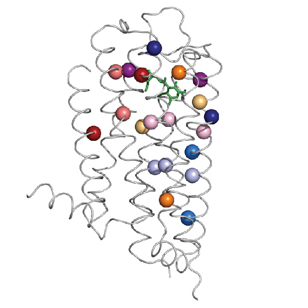
A high throughput microscopy technique enabled Baylor College of Medicine researchers to identify a new modulator of estrogen receptor alpha that they called UBR5. It is a highly amplified gene in many tumors, including breast, prostate and ovarian cancer.

With high throughput microscopy and automated image analysis, researchers can evaluate many mechanistic measures under several hundred experimental conditions simultaneously, said Dr. Michael Mancini, professor of molecular and cellular biology at Baylor and director of the integrated Microscopy Core, in a report that appears online in the journal Oncogene.
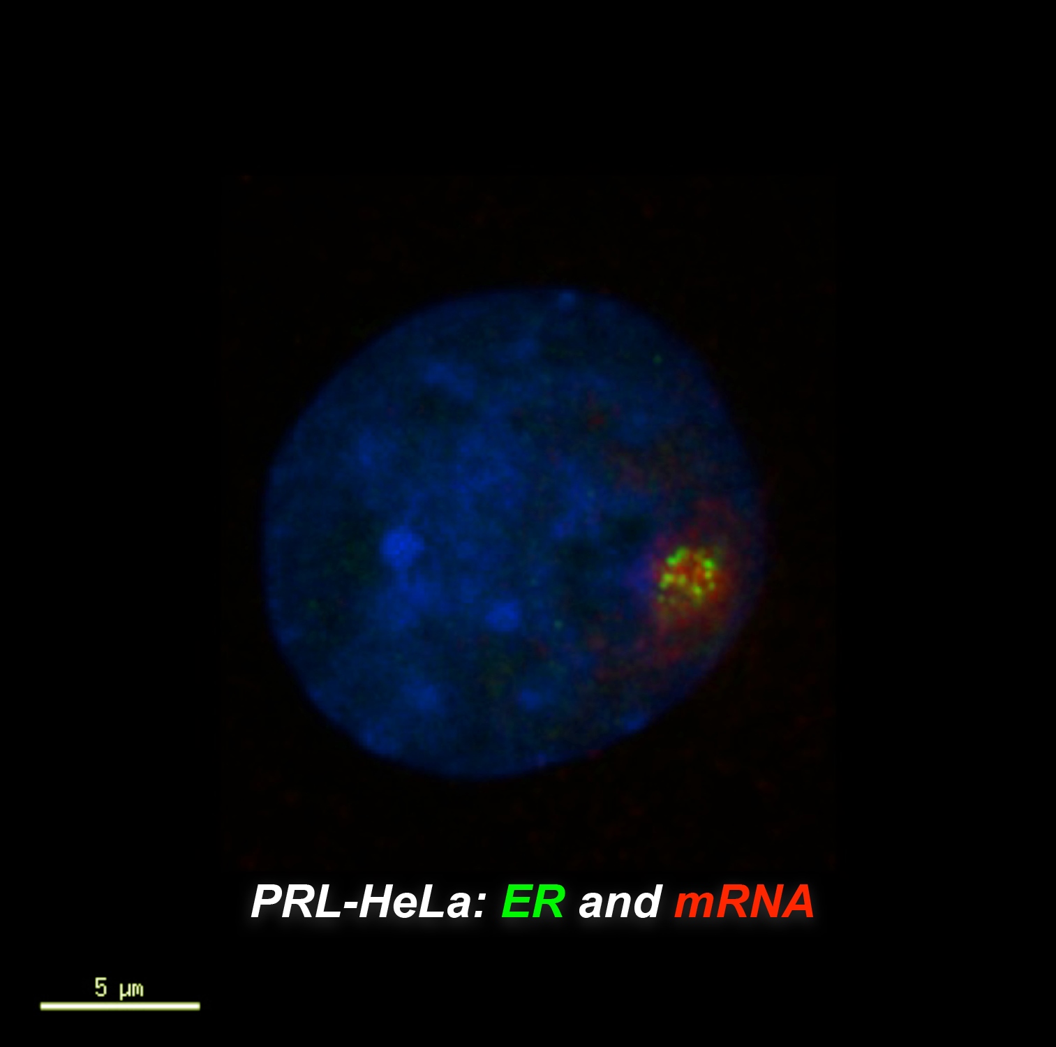
Using siRNA (a gene-silencing technique) for almost 300 different transcription modulators (proteins that govern transcription factor activity) and a special estrogen-responsive biosensor cell line platform, they were able to perform many measures at the same time, including protein levels, DNA binding, chromatin (the building material of chromosomes) modeling, transcription, protein interactions and toxicity.
More than 600,000 images and 45 million data points
Mancini estimates that the project involved more than 600,000 images and 45 million data points.

Michael J. Bolt, a graduate student in Mancini’s laboratory, was first author of the report and performed much of the actual microscopy and analysis.
Dr. Fabio Stossi, an assistant professor in the department of molecular and cellular biology, said, “The highly customized cell platform that has evolved over several years was essential for this project. We’ve identified a novel protein that has some potential for understanding ovarian and some forms of breast cancer.”
New technique
High throughput microscopy is a relatively new technique that involves taking many images and evaluating them via computer for automated data mining. When combined with specifically-engineered cell lines and screening resources, the approach is remarkably powerful.
“In some ways, now, it’s a lot simpler than you think,” said Bolt. “Most of the experimental process is performed robotically. You have to know how it works so that you can tell the robots what to do.”
The appropriate siRNAs and transfection reagents are robotically-deposited into 384 well plates, followed by seeding of the engineered cell lines that facilitate visualization of transcription. Then, plates are loaded onto a recently-acquired high-speed state-of-the-art microscope that automatically takes all the photos. Then, Bolt used custom in-house designed software to analyze all the photos.
Massive spreadsheet
“You plug it in and you get a massive Excel spreadsheet. These are ‘Big Data’ experiments” said Bolt.
“We are quantitatively visualizing all of the engineered transcription loci in every cell,” said Mancini. “Did it affect DNA binding? Promoter loading? Chromatin changes? Transcriptional output? We are collecting much more mechanistic content now using a single integrated assay.”
Others who took part in this work include Austin M. Callison, Maureen G. Mancini and Radhika Dandekar, all of Baylor.
Funding for this work came from the Keck Foundation, the John S. Dunn Gulf Coast for Chemical Genomics, the Dan L Duncan Baylor Cancer Center , the Baylor Center for Reproductive Biology, the Keck Center National Library to Medicine Training Program in Biomedical Informatics of the Gulf Coast Consortia National Library of Medicine [ Grant T15LM007093], the Diana Helis Henry Medical Research Foundation through its direct engagement in the continuous active conduct of medical research in conjunction with Baylor College of Medicine and the Cancer Program and the Integrated Microscopy Core at Baylor College of Medicine with funding from the NIH (Grants HD007495, DK56338, and CA125123). — Ruth SoRelle, M.P.H.